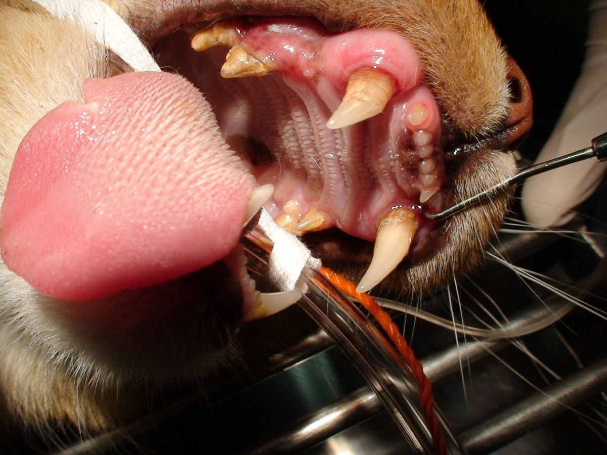
Dental Pathology
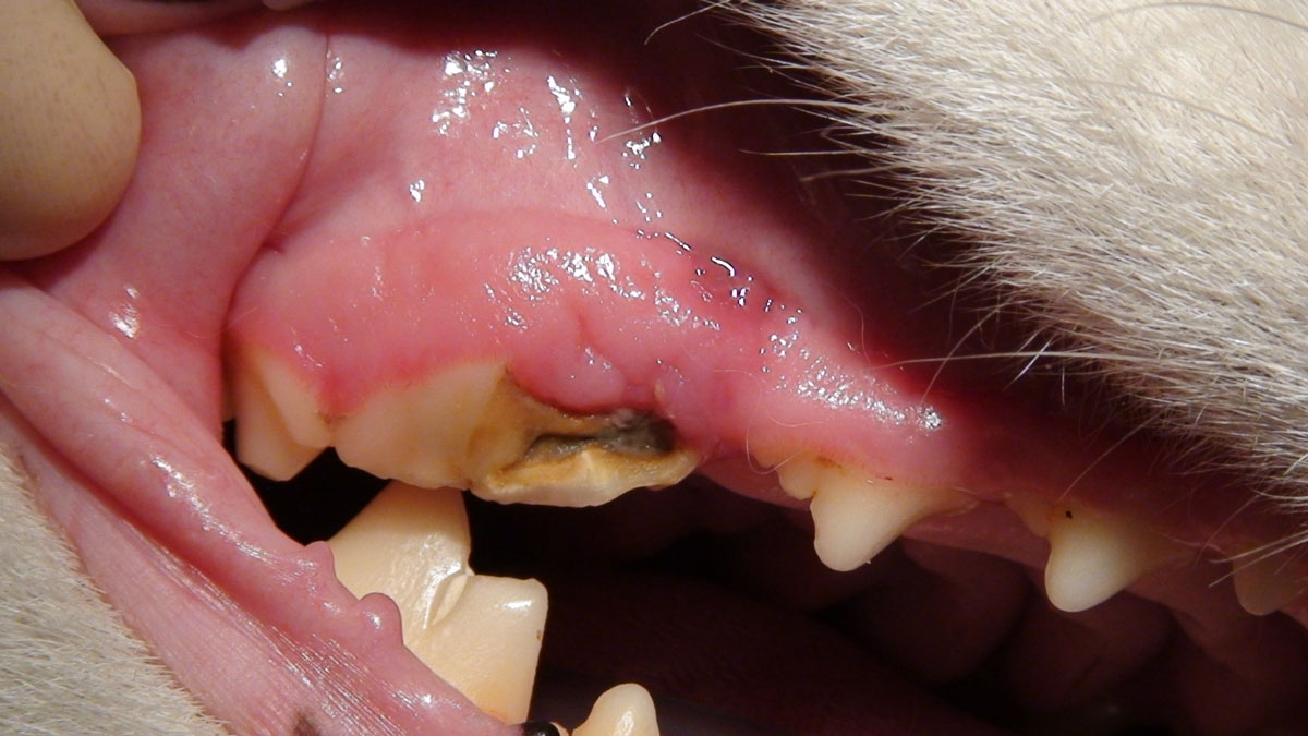
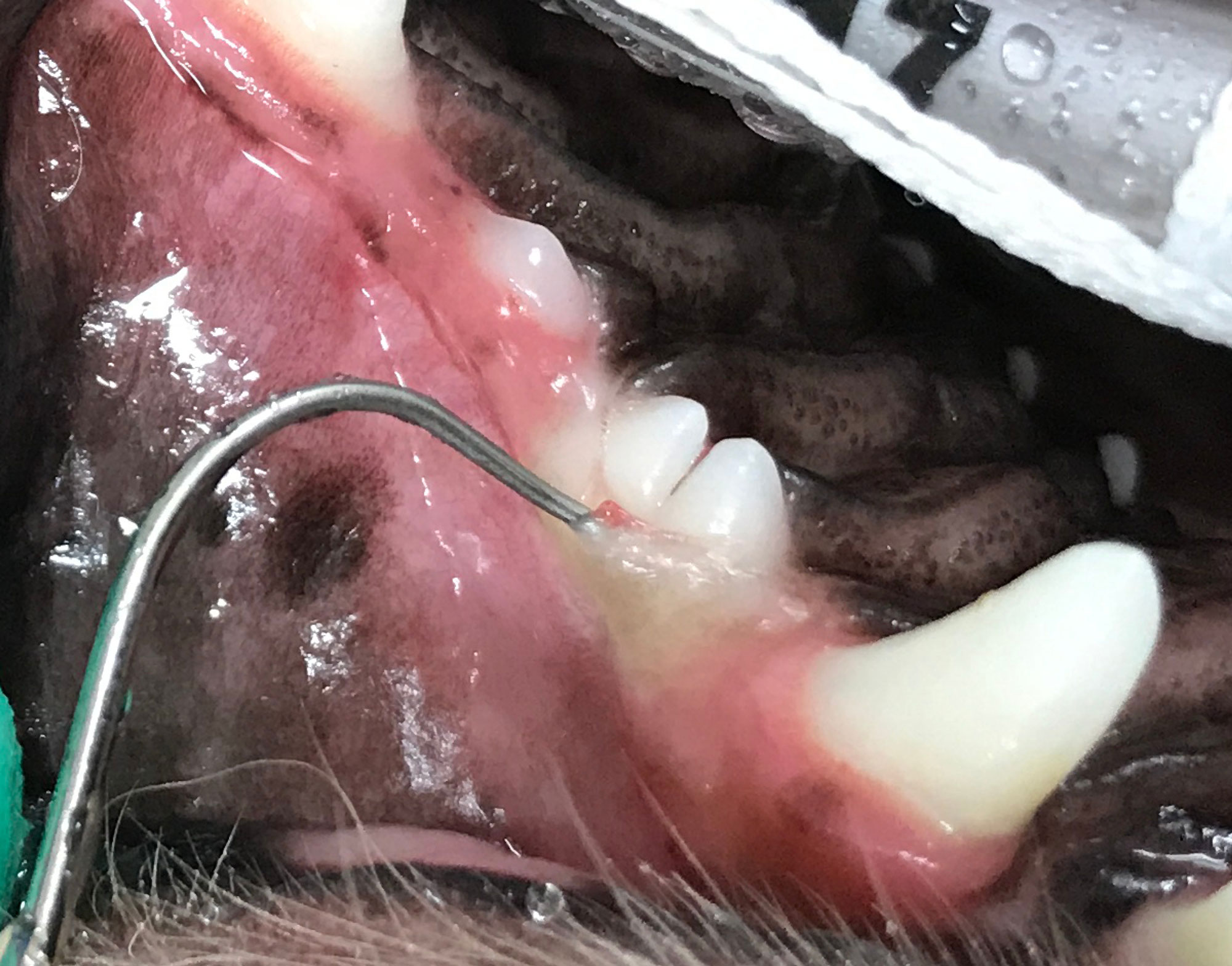
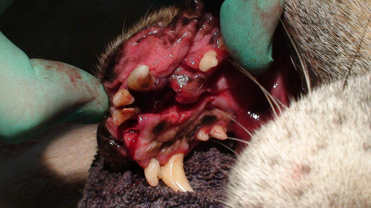
Near pulp exposure
The healthy tooth is composed of two major parts: the supra-gingival portion that extends above the gingiva and is grossly visible, termed the crown, and the sub-gingival portion that extends beneath the gingiva, termed the root. A central cavity is located within the tooth, termed the pulp canal (located in the root segment) and pulp chamber (located in the crown segment). The pulp, which consists of nerves, blood vessels and connective tissues, is located within the pulp canal and chamber. It provides nutrition and sensation to the tooth. The pulp is surrounded and therefore protected by two hard layers. An inner layer composed of dentine, which is found in both the crown and root, and an outer layer of enamel and cementum. The crown is covered in enamel. The root has an outer covering of cementum. The point where the crown and root meet is termed the cemento-enamel junction. Loss of tooth structure resulting in only a thin layer of dentine covering over the pulp may produce a near pulp exposure. Treatment is aimed as indirect pulp capping to cover any exposed dentinal tubules and prevent ingress of bacteria to the pulp.
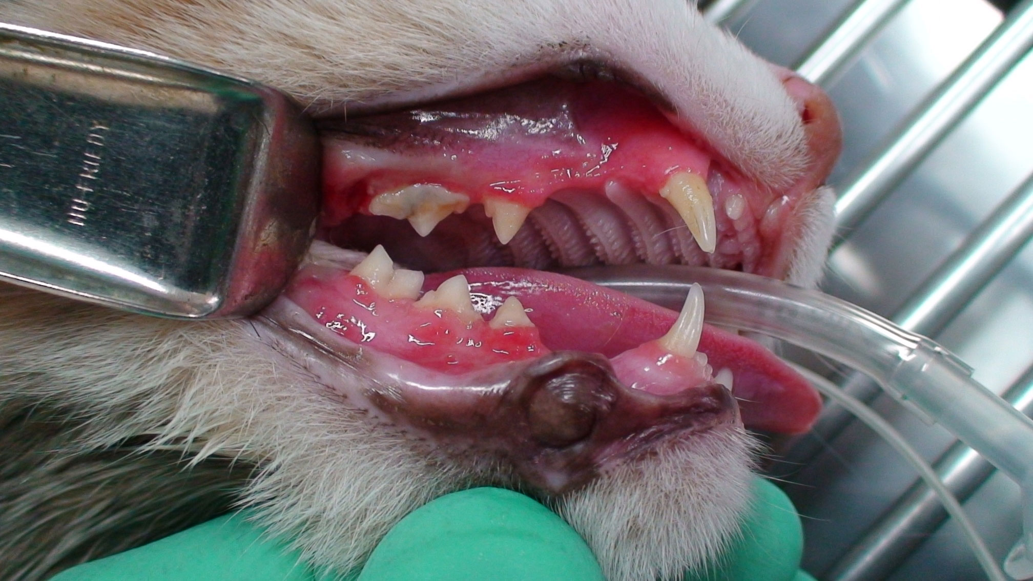
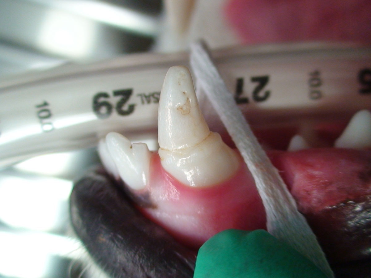
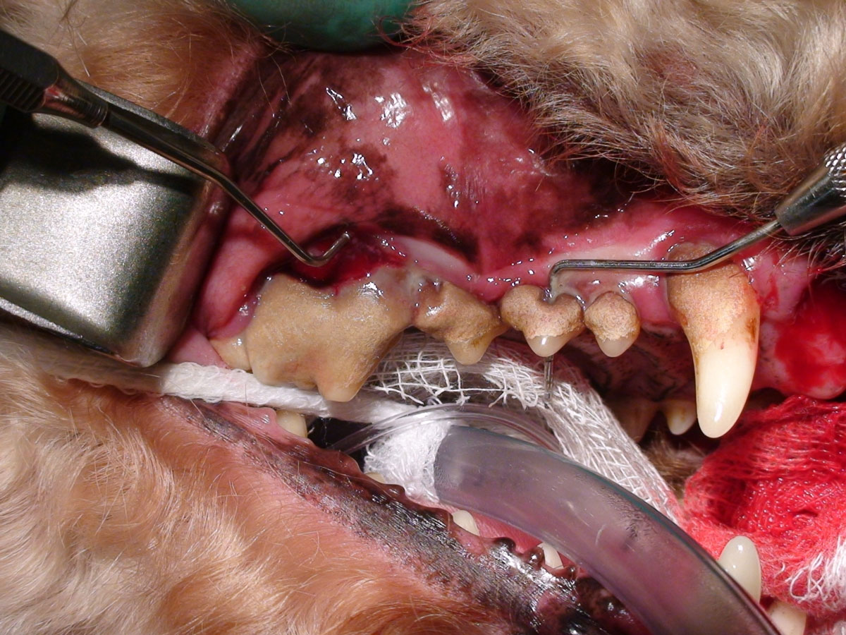
Uncomplicated crown-root facture
The healthy tooth is composed of two major parts: the supra-gingival portion that extends above the gingiva and is grossly visible, termed the crown, and the sub-gingival portion that extends beneath the gingiva, termed the root. A central cavity is located within the tooth, termed the pulp canal (located in the root segment) and pulp chamber (located in the crown segment). The pulp, which consists of nerves, blood vessels and connective tissues, is located within the pulp canal and chamber. It provides nutrition and sensation to the tooth. The pulp is surrounded and therefore protected by two hard layers. An inner layer composed of dentine, which is found in both the crown and root, and an outer layer of enamel and cementum. The crown is covered in enamel. The root has an outer covering of cementum. The point where the crown and root meet is termed the cemento-enamel junction. An UCRF is one which removes enamel, dentine and cementum and does not expose the pulp. As such the dentinal tubules are exposed if the dentine is fractured and treatment is aimed at removing rough edges, decreasing pulpitis, sealing the dentinal tubules and restoring normal anatomy where possible and practical.
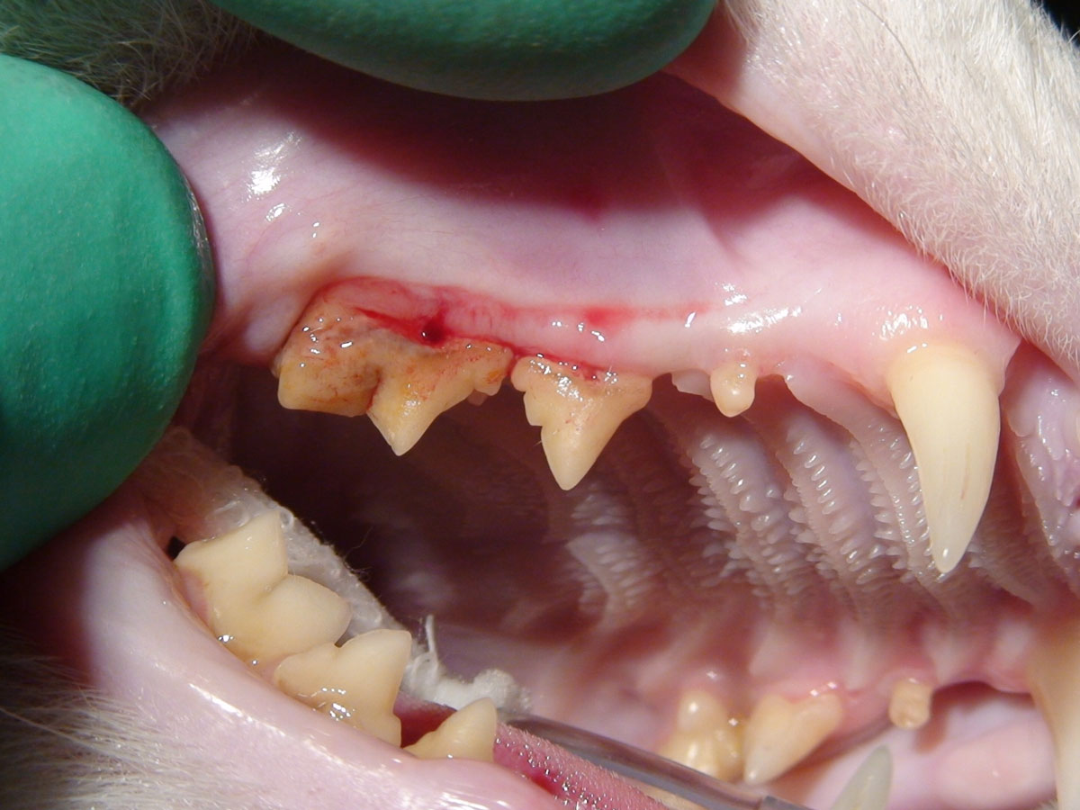
Uncomplicated crown fracture
The healthy tooth is composed of two major parts: the supra-gingival portion that extends above the gingiva and is grossly visible, termed the crown, and the sub-gingival portion that extends beneath the gingiva, termed the root. A central cavity is located within the tooth, termed the pulp canal (located in the root segment) and pulp chamber (located in the crown segment). The pulp, which consists of nerves, blood vessels and connective tissues, is located within the pulp canal and chamber. It provides nutrition and sensation to the tooth. The pulp is surrounded and therefore protected by two hard layers. An inner layer composed of dentine, which is found in both the crown and root, and an outer layer of enamel and cementum. The crown is covered in enamel. The root has an outer covering of cementum. The point where the crown and root meet is termed the cemento-enamel junction. A UCF is one which removes enamel, or enamel and dentine, is a fracture of the crown and does not expose the pulp. As such the dentinal tubules are exposed if the dentine is fractured and treatment is aimed at removing rough edges, decreasing pulpitis, sealing the dentinal tubules and restoring normal anatomy where possible and practical.
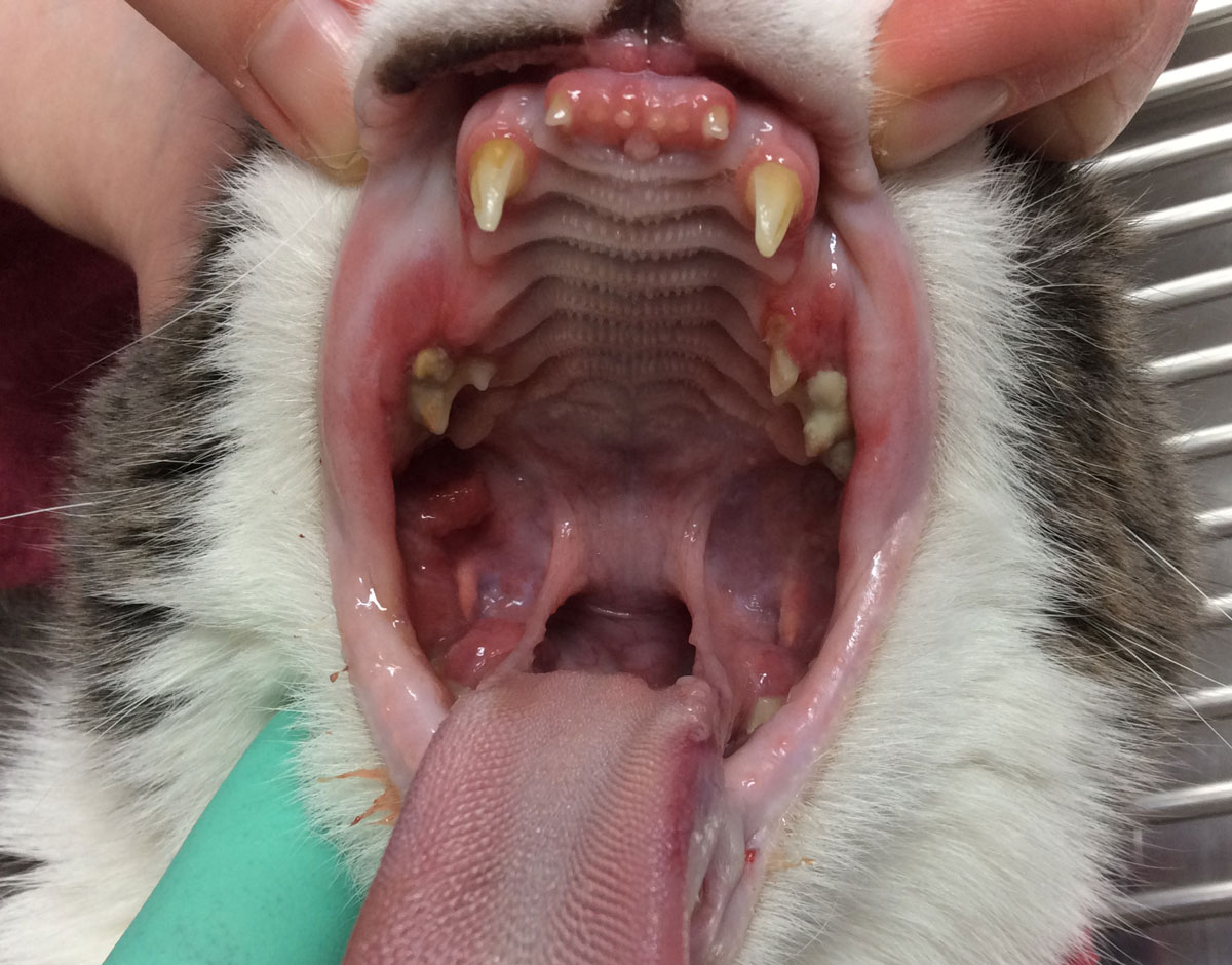
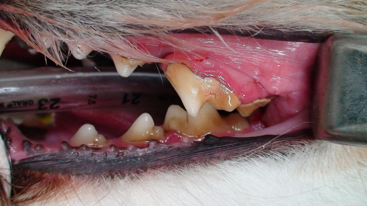
Tooth resorption
Classification of Tooth Resorption
- Stage 1 (TR 1): Mild dental hard tissue loss (cementum or cementum and enamel).
- Stage 2 (TR 2): Moderate dental hard tissue loss (cementum or cementum and enamel with loss of dentin that does not extend to the pulp cavity).
- Stage 3 (TR 3): Deep dental hard tissue loss (cementum or cementum and enamel with loss of dentin that extends to the pulp cavity), most of the tooth retains its integrity.
- Stage 4 (TR 4): Extensive dental hard tissue loss (cementum or cementum and enamel with loss of dentin that extends to the pulp cavity), most of the tooth has lost its integrity. (TR4a) Crown and root are equally affected, (TR4b) Crown is more severely affected than the root, (TR4c) Root is more severely affected than the crown.
- Stage 5 (TR 5): Remnants of dental hard tissue are visible only as irregular radiopacities, and gingival covering is complete.
This classification is based on the assumption that tooth resorption is a progressive condition.
Treatment of tooth resorption is to remove the pain and any associated infection. In cats with external resorption this involves obtaining radiographs and determining whether there is ankylosis. Teeth that have a healthy periodontal liagament should be extracted completely, whereas ankylosed teeth may be reduced in height by crown amputation and covered by suturing the gingival over the remaining root. Teeth undergoing internal resorption my either be extracted or have a root canal procedure performed.
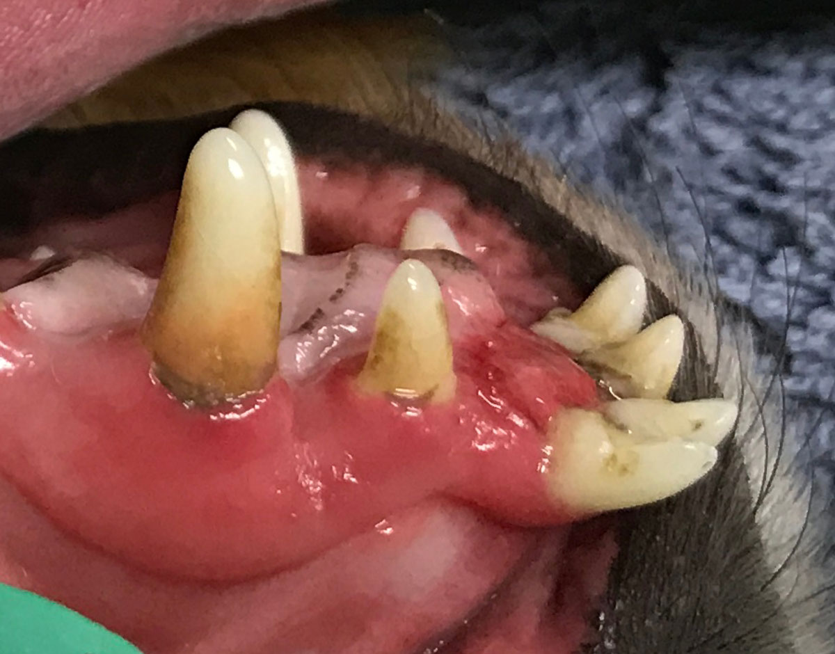
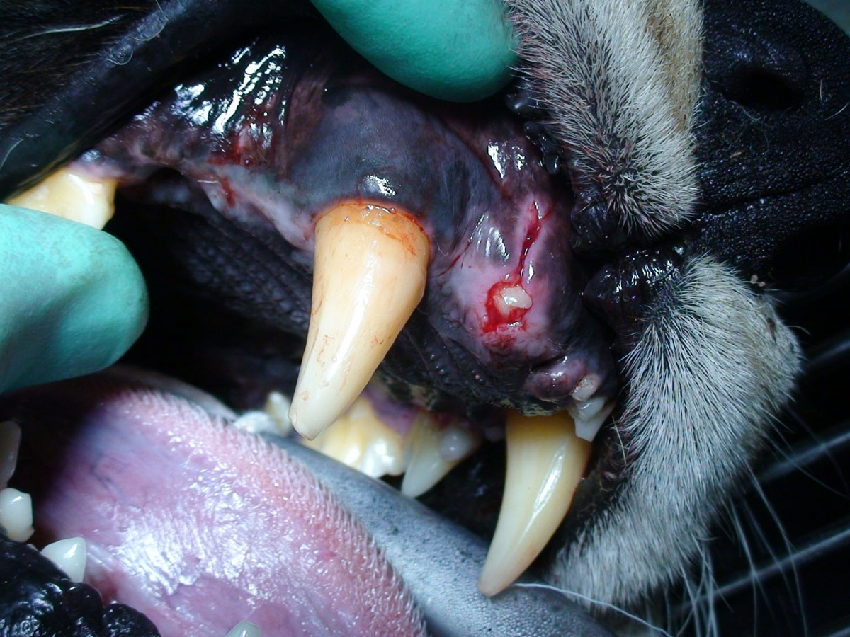
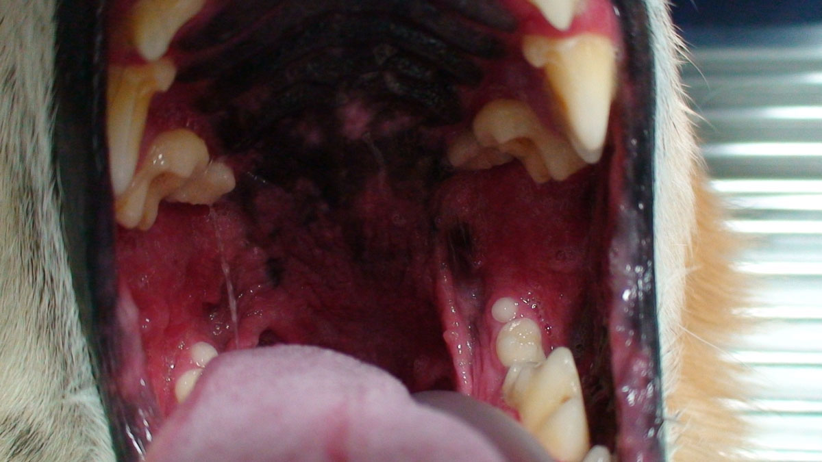
Retained tooth root
The abnormal present of a tooth root without an associated crown. This may be observed covered by gingival and found on radiographs, or penetrating the gingival following inadequate extraction or loss of crown after trauma or resorption. Radiographs are mandatory to determine the degree of pathology.
Pathology Treatment:
A self resorbing root causing no obvious inflammation or pain may be monitored, whereas a partially erupted root demonstrating gingival inflammation and pain should be extracted.
Abrasion
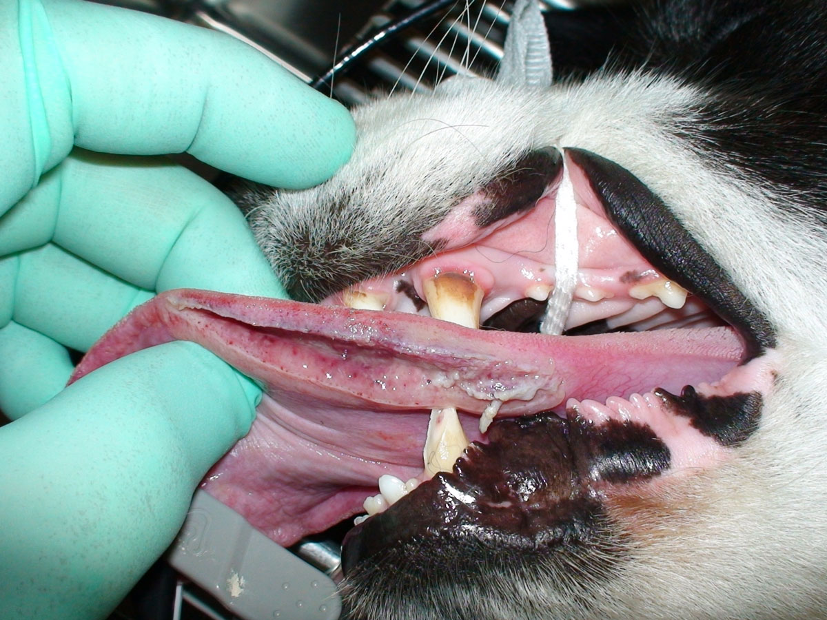
Excessive wear from friction of an externally applied force, such as rock, bone or toothbrushing may result in loss of enamel and/or dentine. When the friction is light or gradual, the tooth responds by the odontoblasts producing tertiary dentine to seal and protect the underlying pulp. Tertiary dentine is often observed as a honey brown discolouration of the tooth, is smooth on probing and appears schlerotic and disorganized on radiograph. If the wear is rapid, or if the odontoblasts do not produce adequate tertiary dentine to ensure the pulp remains covered, the pulp canal becomes open and the pulp exposed to the oral environment, leaving it open for infection and subsequent pulpitis.
Attrition
There is a normal amount of wear of the tooth surface from normal mastication, the force of tooth against tooth contact of molars, malocclusions of other teeth and chewing the hair coat, this is termed attrition. When the wear is light or gradual, the tooth responds by the odontoblasts producing tertiary dentine to seal and protect the underlying pulp. Tertiary dentine is often observed as a honey brown discolouration of the tooth, is smooth on probing and appears schlerotic and disorganized on radiograph. If the wear is rapid, or if the odontoblasts do not produce adequate tertiary dentine to ensure the pulp remains covered, the pulp canal becomes open and the pulp exposed to the oral environment, leaving it open for infection and subsequent pulpitis.
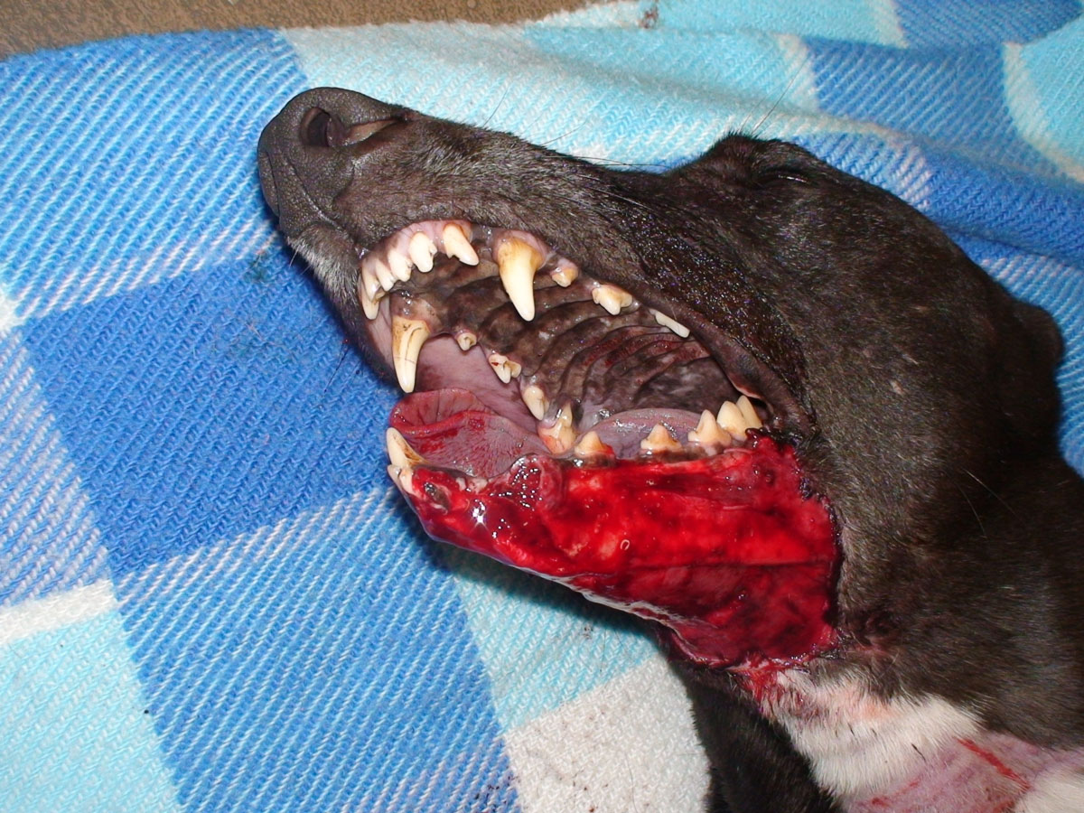
Avulsed Tooth
Complete displacement of the tooth from the socket due to trauma or removal of a tooth often due to trauma leaving an empty socket. Treatment for a permanent tooth requires immediate replantation, splinting and often future root canal treatment.
Bigeminy
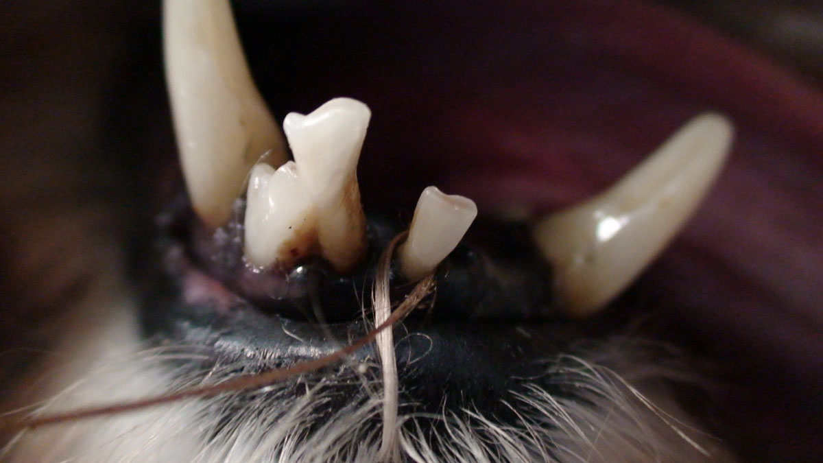
Bigeminy includes both tooth fusion and tooth germination. Fusion arises through union of two normally separated tooth germs, and depending upon the stage of development of the teeth at the time of union, it may be either complete or incomplete. Germination arises when two teeth develop from one tooth bud, it appears that the dog or cat has an extra tooth, although they have a normal number of tooth roots.
Canine bigeminy example one
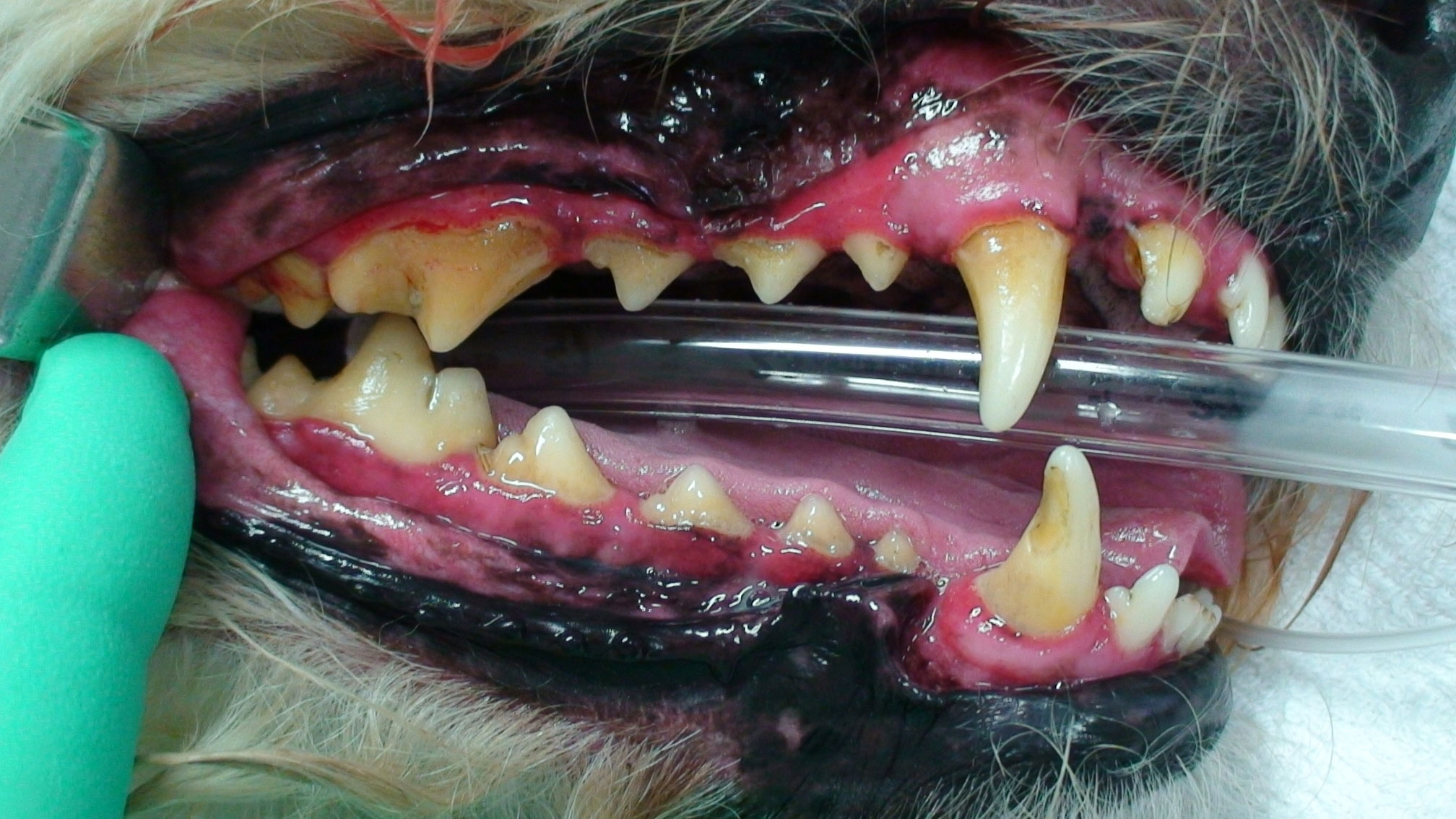
Calculus Index
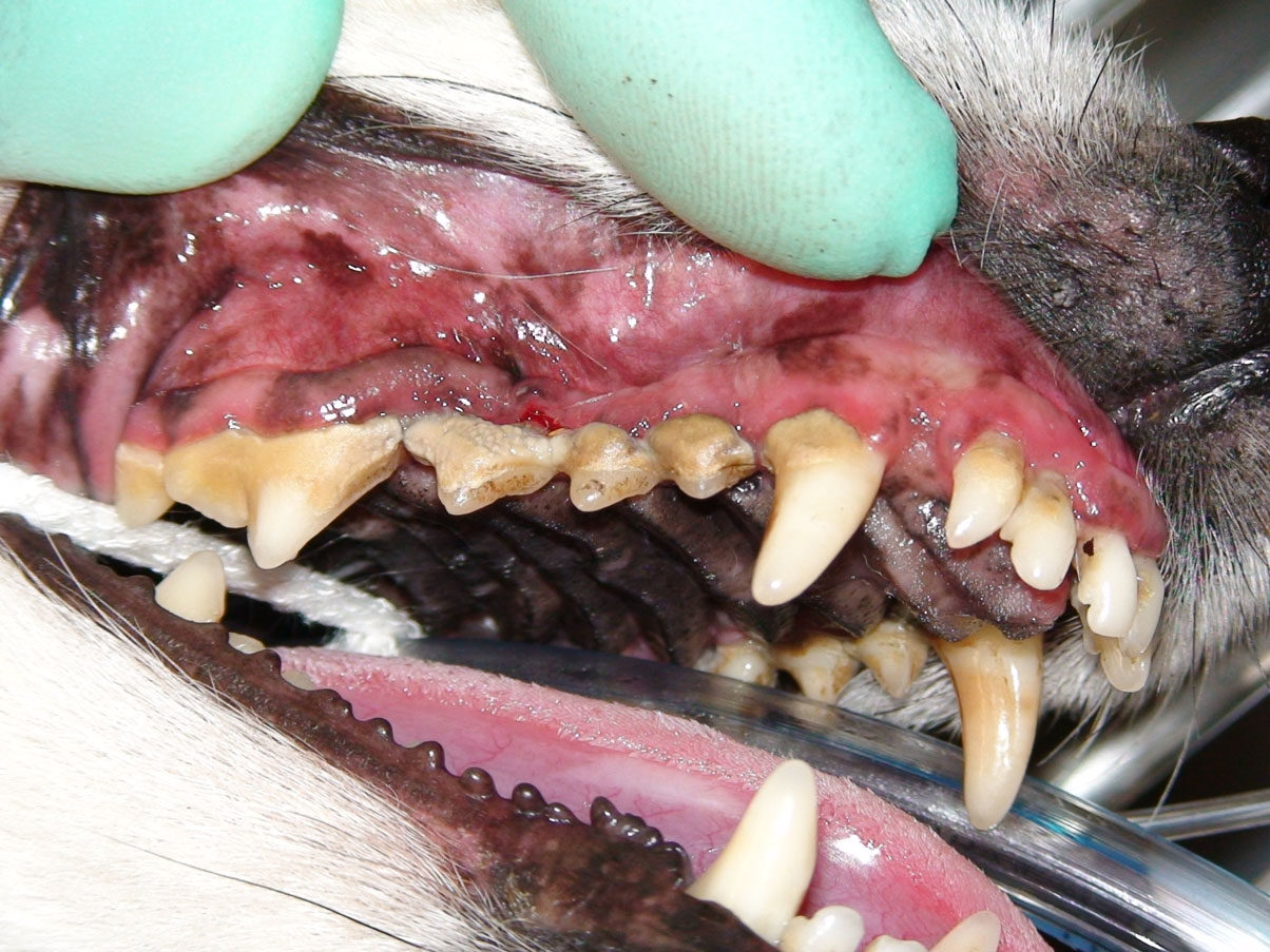
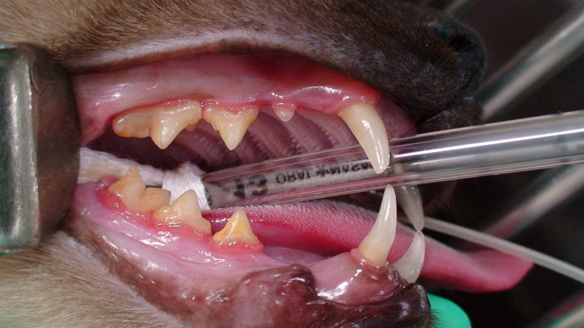
Calculus is mineralized plaque that accumulates on hard tooth surfaces, both supra- and sub-gingival. Calculus accumulates faster on the buccal surface of teeth, in particular adjacent to the maxillary 4th premolars and parotid and zygomatic salivary duct openings. Whilst calculus does not directly result in periodontal disease, it certainly aids in the accumulation of plaque, acts as a local irritant and prevents healing of subgingival tissues. The treatment aim is to thoroughly remove all calculus form the tooth surface. Calculus accumulation can be classified by coverage of tooth surface area where the Calculus Index (CI #) refers to:
Pathology Treatment:
The treatment aim is to thoroughly remove all calculus form the tooth surface.
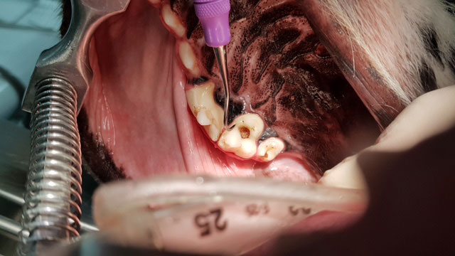
Caries
A demineralisation of the enamel as a result of bacteria producing acids are most commonly located on the flat occlusal surfaces of the maxillary 1st and 2nd molars and the mandibular 1st, 2nd and 3rd molars in dogs. An incipient carious lesion of enamel often appears as a chalky white spot. A carious dental lesion can be differentiated from staining or tooth resorption as an explorer will often catch on withdrawal after probing and feel often stick in the carious dentin. Compare this to healthy dentin where the explorer can be drawn across it with scratchy sound and feel. Treatment is aimed as drilling out the carious dentine until sound dentine remains and a restorative placed.
Pathology Treatment:
Treatment is aimed as drilling out the carious dentine until sound dentine remains and a restorative placed.
Complicated crown-root fracture
The healthy tooth is composed of two major parts: the supra-gingival portion that extends above the gingiva and is grossly visible, termed the crown, and the sub-gingival portion that extends beneath the gingiva, termed the root. A central cavity is located within the tooth, termed the pulp canal (located in the root segment) and pulp chamber (located in the crown segment). The pulp, which consists of nerves, blood vessels and connective tissues, is located within the pulp canal and chamber. It provides nutrition and sensation to the tooth. The pulp is surrounded and therefore protected by two hard layers. An inner layer composed of dentine, which is found in both the crown and root, and an outer layer of enamel and cementum. The crown is covered in enamel. The root has an outer covering of cementum. The point where the crown and root meet is termed the cemento-enamel junction. A CCRF is one which removes enamel and dentine from the crown, cementum and dentine from the root and does expose the pulp. As such the dentinal tubules and the pulp canal are exposed leading to pulpitis and pain. Treatment is aimed at removing rough edges, decreasing pulpitis, preventing periapical abscessation or treating it and restoring normal anatomy where possible and practical. Where the fracture is less than 48 hours duration, direct pulp capping is preferred, whereas older pulp exposes require root canal treatment or extraction.
Dentigerous Cyst
Dentigerous cysts (follicular cysts) are the most common odontogenic cysts in dogs and cats are associated with the crown of an unerupted tooth, usually a permanent tooth with the mandibular 1st premolar often over-represented. They arise from the epithelial remnants of the enamel organ or the reduced enamel epithelium that surrounds the crown during odontogenesis. All missing teeth should be radiographed, and the cyst requires surgical removal of the epithelial lining and removal of the tooth.
Enamel hypocalcification or hypoplasia
Enamel Hypocalcification or Enamel Hypoplasia: The basic building block of the crown enamel is the enamel rod, which is a column of enamel extending from the dentino-enamel junction to the coronal surface of the tooth. Enamel is produced by ameloblasts, initially mineraliasation occurs where the enamel rod matrix is laid down, followed by maturation, where the crystals grow in size and become tightly packed together. If the crystals fail to grow to full size, the rods will be poorly calcified, a condition termed enamel hypocalcification, and will often appears as a pit with dentine exposure. When the lesion is probed with an explorer it appears hard but brittle. Enamel hypoplasia is a defect of the tooth crown where the enamel is deficient in thickness thus is thin.
Furcation
The furcation is the point where the roots separate in a multi-rooted tooth and is often also considered the space between the tooth roots. Furcation involvement and problems occur with attachment loss in this area due to food retention, plaque retention, increased attachment loss, carious lesions. Detection of furcation exposure is determined by use of a probe and radiographs. Exposure exists when a periodontal probe extends into or through the space. Furcation involvement can be divided into the following classifications:
- Stage 0 (F0) – No furcation involvement.
- Stage 1 (F1) – a probe extends less than half way under the crown in any direction of a multirooted tooth with attachment loss.
- Stage 2 (F2) – a probe extends greater than half way under the crown of a multirooted tooth with attachment loss but not through to the other side.
- Stage 3 (F3) – a probe extends under the crown of a multirooted tooth, through from one side of the furcation to exit the other side.
Pathology Treatment:
Add treatment aims and link to full treatment page for members
Gingivitis
The gingivitis index is designed to give a quantitative measure of the degree of inflammation of the gingival associated with a particular tooth. Once plaque accumulates on the tooth surface, initial gingivitis begins in 2-4 days as the host mounts an immune response to the invading bacteria. The intensity of gingivitis is determined by the quantity and quality of the microorganisms present. When gingivitis is manifest clinically, there is generally a plaque layer of 400um thick. Once this occurs, the host’s response is to produce an exudate, elevated sulcus fluid flow and leukocyte (PMN) infiltration. The Gingivitis Index (GI #) is classified as follows:
GI 0 normal healthy gingiva with sharp non inflamed margins.
GI 1 marginal gingivitis with minimal inflammation and oedema at the free gingiva. No bleeding on probing.
GI 2 moderate gingivitis with an increase in inflammation and oedema throughout free gingivia and delayed bleeding upon probing.
GI 3 advanced gingivitis with inflammation clinically reaching the mucogingival junction usually with ulceration. Immediate bleeding on probing.
Pathology Treatment:
Add treatment aims
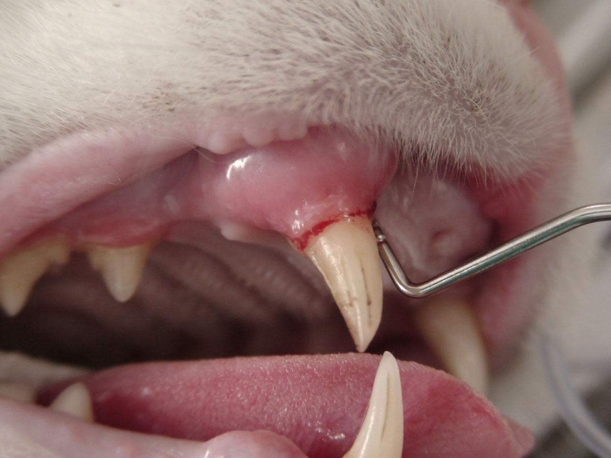
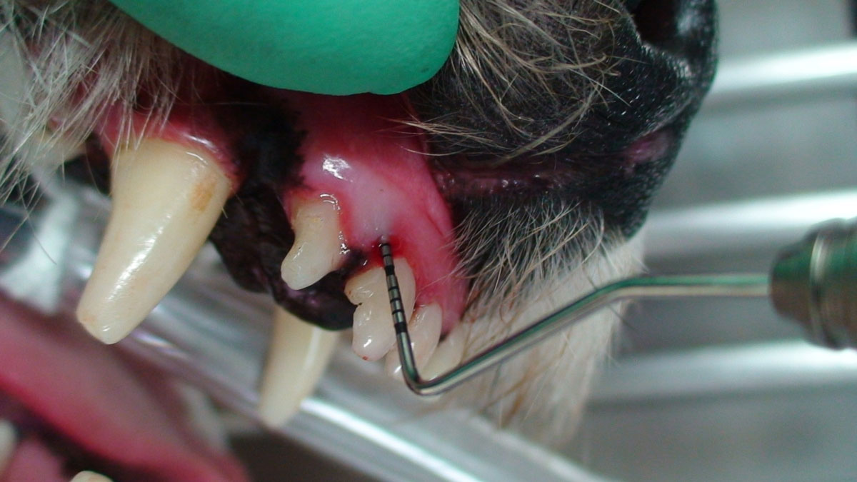
Gingival recession
Gingival margin recession is observed as the retreat in an apical direction of the buccal periodontium. Recession of the gingival margin exposes the cementum and in multiple rooted teeth, the furcation. It is common for the tooth to maintain a normal periodontal sulcus depth, so accurate attachment loss should be measured from the gingival margin to the cement-enamel junction (CEJ).
Pathology Treatment:
Treatment aims to remove any endotoxins and accumulation form the tooth root, clean the sulcus or pocket and seal the exposed dentinal tubules.
Intrinsic Stain
Intrinsic staining or discolouration within the tooth is commonly caused by
1. Haemorrhage following trauma with loss of dentinal odontoblasts and movement of erythrocytes into the empty tubules,
2. Discolouration by medications or drugs such as tetracyclines incorporated into the tooth when it is formed,
3. Other medications that bind to the dentine.
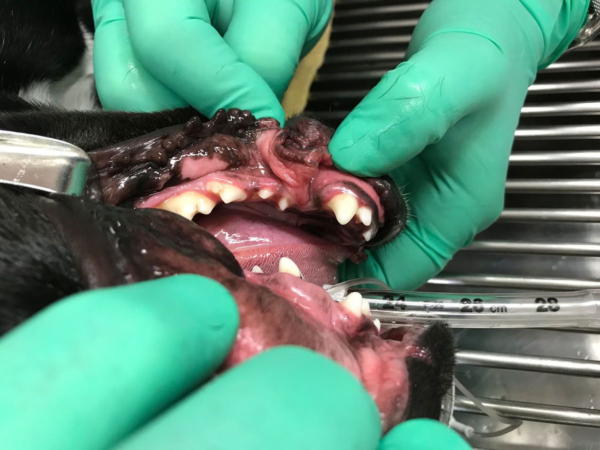
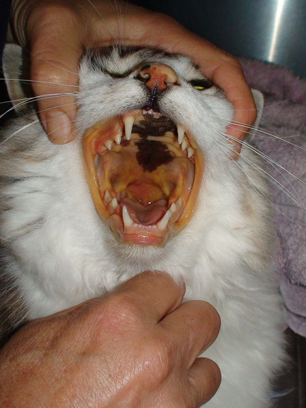
Mesially Deviated Canine
The maxillary canine tooth when mesially deviated may be caused by a genetic cause, such as in Shetland Sheepdogs, or malocclusion of the mandible which results in the mandibular canine traumatising the maxillary canine or trauma which moves the permanent tooth bud. The mesial position of the maxillary tooth often closes the diastema and forces the mandibular canine buccally. Many treatment options are available.
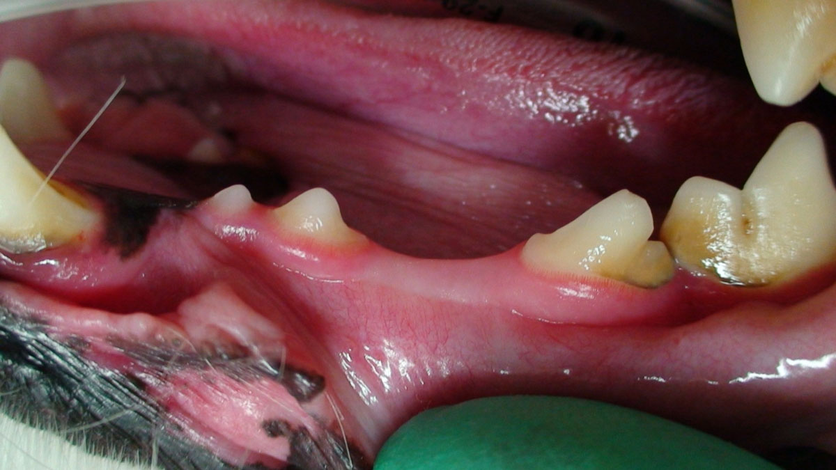
Missing tooth
An absent or missing tooth is recorded when visually there is no tooth present. The ideal treatment involves initially obtaining a radiograph to determine if the tooth is actually absent, whether the crown has been lost and the roots remain, or the tooth has not erupted and is retained. Treatment options are then considered.
Pathology Treatment:
The ideal treatment involves initially obtaining a radiograph to determine if the tooth is actually absent, whether the crown has been lost and the roots remain, or the tooth has not erupted and is retained. Treatment options are then considered.
Mobility
The mobility of a tooth is a critical diagnostic and prognostic tool. The degree of normal physiological movement is limited to the width and elasticity of the periodontal ligament. Abnormal, or pathological, movement increases with loss of supporting tissues (alveolar bone, periodontal ligament) or following trauma. The degree of movement can be classified as follows:
- Stage 0 (M0) – Physiologic mobility up to 0.2 mm.
- Stage 1 (M1) – mobility is increased in any direction other than axial over a distance of more than 0.2 mm and up to 0.5 mm.
- Stage 2 (M2) – the mobility is increased in any direction other than axial over a distance of more than 0.5 mm and up to 1.0 mm.
- Stage 3 (M3) – the mobility is increased in any direction than axial over a distance exceeding 1.0 mm or any axial movement.
Pathology Treatment:
The ideal treatment involves initially obtaining a radiograph to determine if the tooth is actually absent, whether the crown has been lost and the roots remain, or the tooth has not erupted and is retained. Treatment options are then considered.
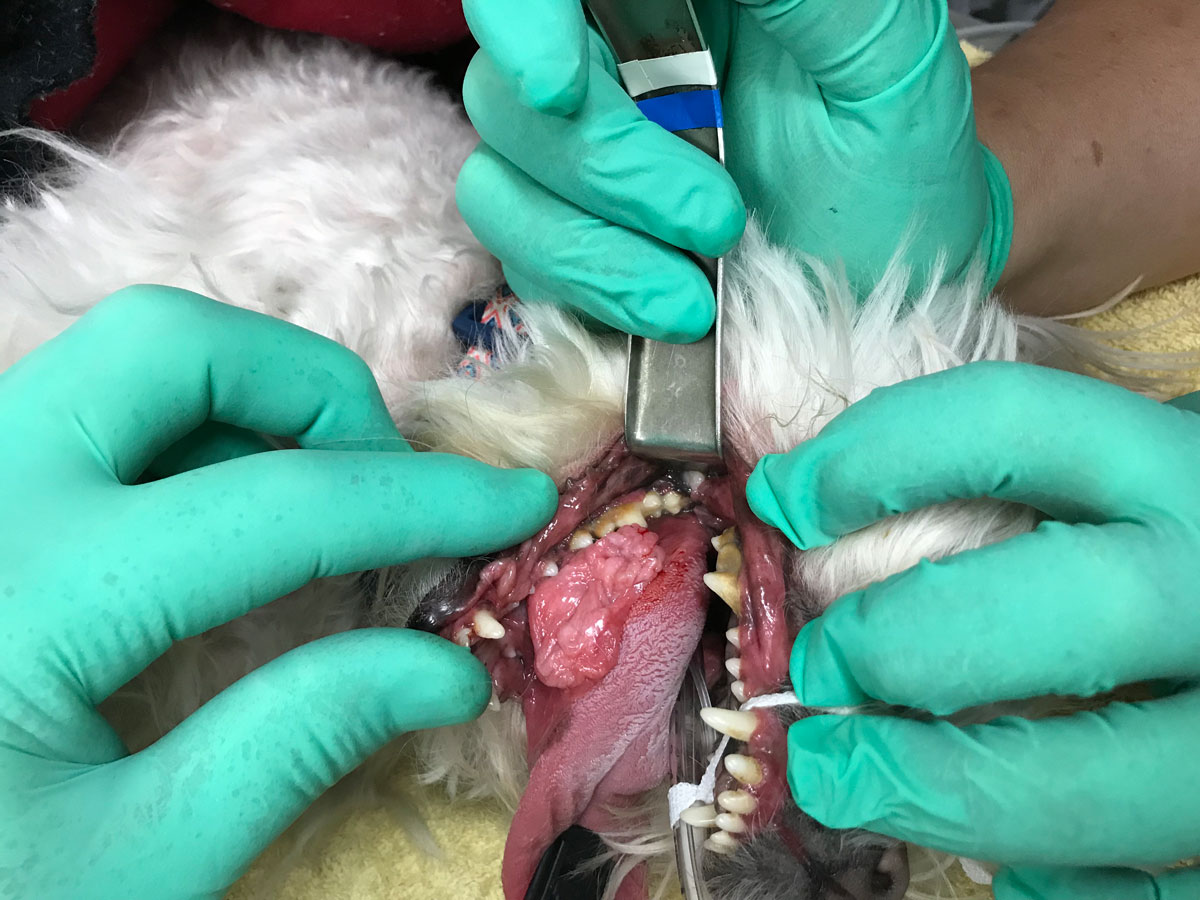
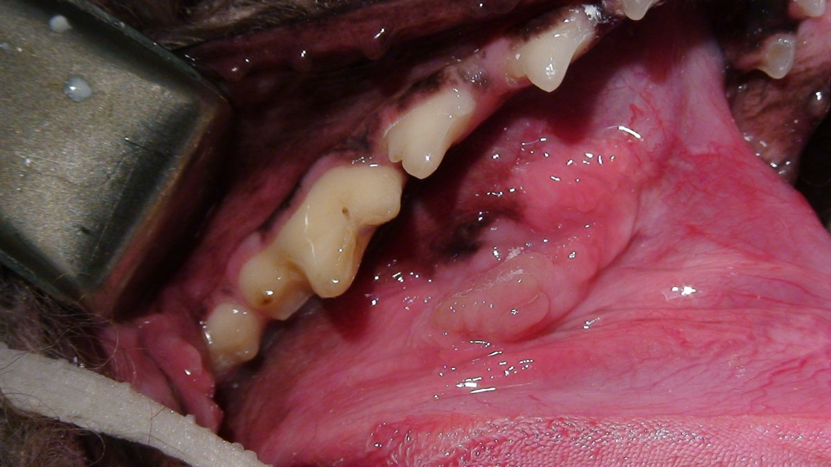
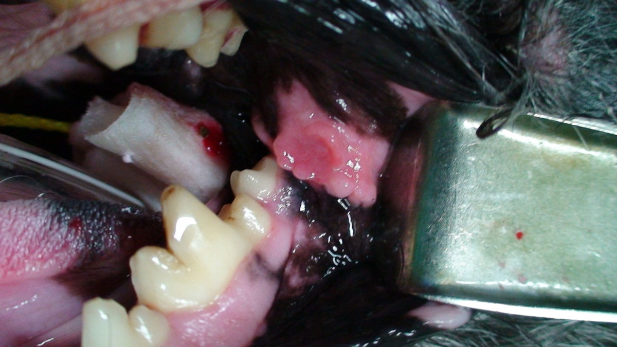
Oral Mass
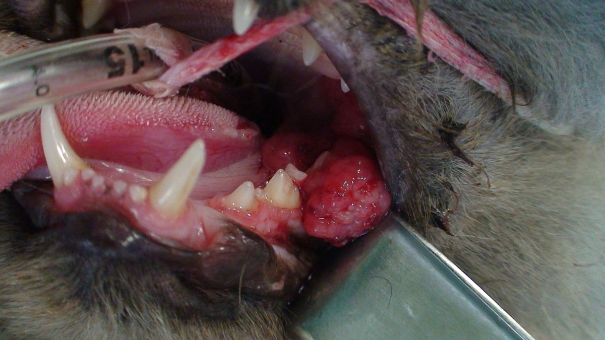
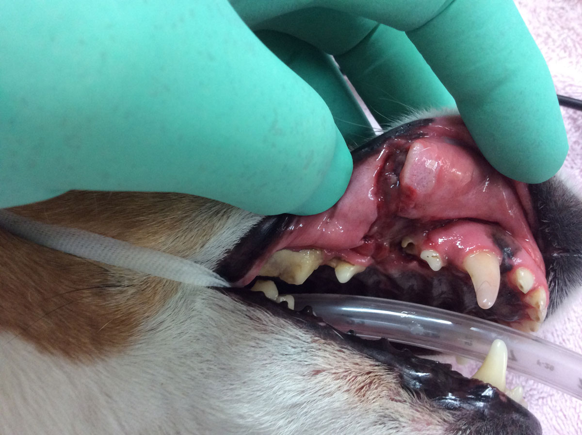
An abnormal tissue growth including both benign and malignant types. Oral tumours are very common in the oral cavity, accounting for 3-12% of all tumours in cats and 6% in dogs. Malignant melanoma and squarmous cell carcinoma are the most common on dogs and squarmous cell carcinoma in cats. Benign oral tumours commonly found include peripheral odontogenic fibroma, ossyfing epulis and acanthomatous ameloblastoma.
Pathology Treatment:
All oral masses should be biopsied and histopathology performed to determine the type od mass and the appropriate treatment.
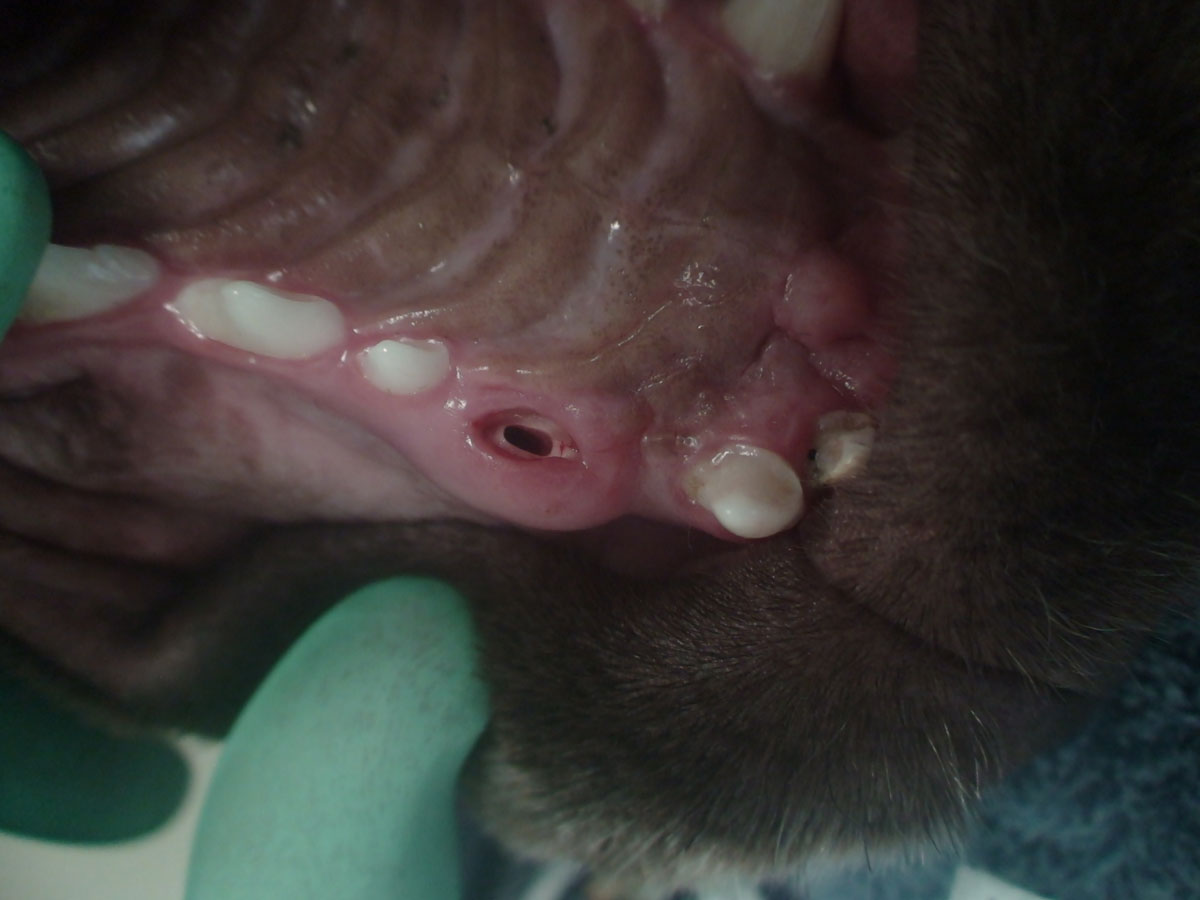
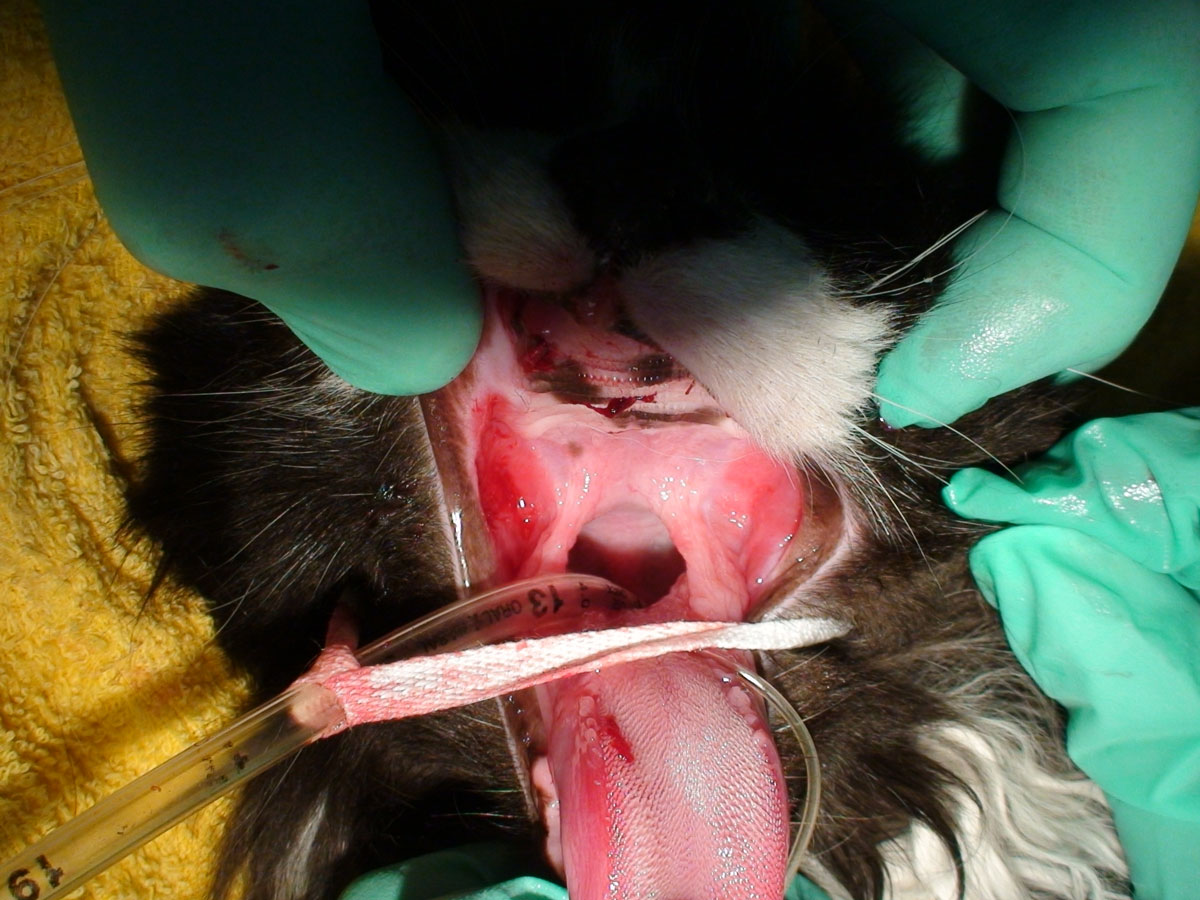
Oronasal fistula
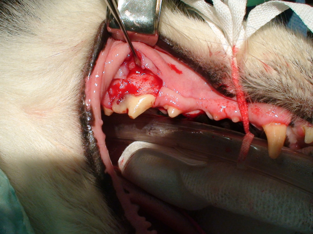
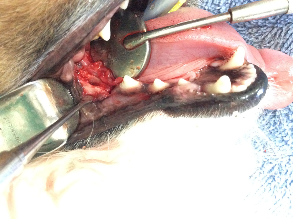
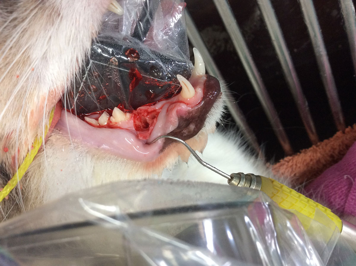
A communication between the oral cavity and the nasal cavity or respiratory tract may be present in any species and adjacent to any tooth. The most common fistula, which be present prior to extraction due to advanced periodontal disease, or occur during extraction, is at the level of the maxillary canine tooth. This is a common site in the Dachshund breed dog. It is not uncommon to observe epistaxis from the side of the fistula during extraction. The goal of treatment is to repair the fistula using sound surgical technique: ensuring there is no tension on the flap, harvesting a large vascular flap, taking large bites of tissue when suturing, confirming knots are secure, not suturing over a void. Either a single or double flap technique may be employed.
Pathology Treatment:
The goal of treatment is to repair the fistula using sound surgical technique: ensuring there is no tension on the flap, harvesting a large vascular flap, taking large bites of tissue when suturing, confirming knots are secure, not suturing over a void. Either a single or double flap technique may be employed.
Periodontal disease index
Whilst a classification of periodontal disease may aid in potential treatment and planning, it is seldom the case that the entire dentition is at the same stage of disease. PDI was originally a useful index for human epidemiological studies, and used six teeth as a representation of the entire mouth. In pets, a percentage attachment loss due to the variation in patient size and thus the depths of pockets can be used as follows:
- PD0 – no attachment loss: normal periodontium.
- PD1 – no attachment loss: gingivitis.
- PD2 – 25% attachment loss: early periodontal disease.
- PD3 – 25 – 50% attachment loss: moderate periodontal disease.
- PD4 – 50% attachment loss: severe periodontal disease.
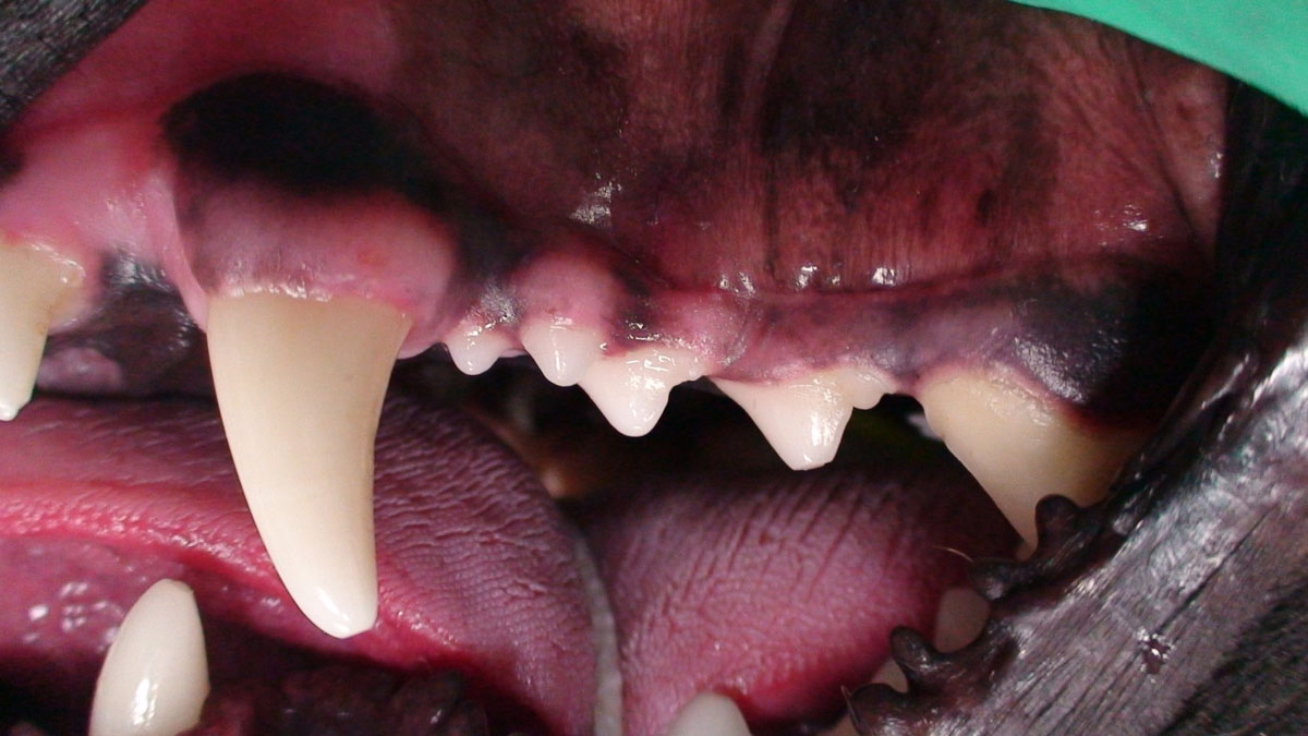
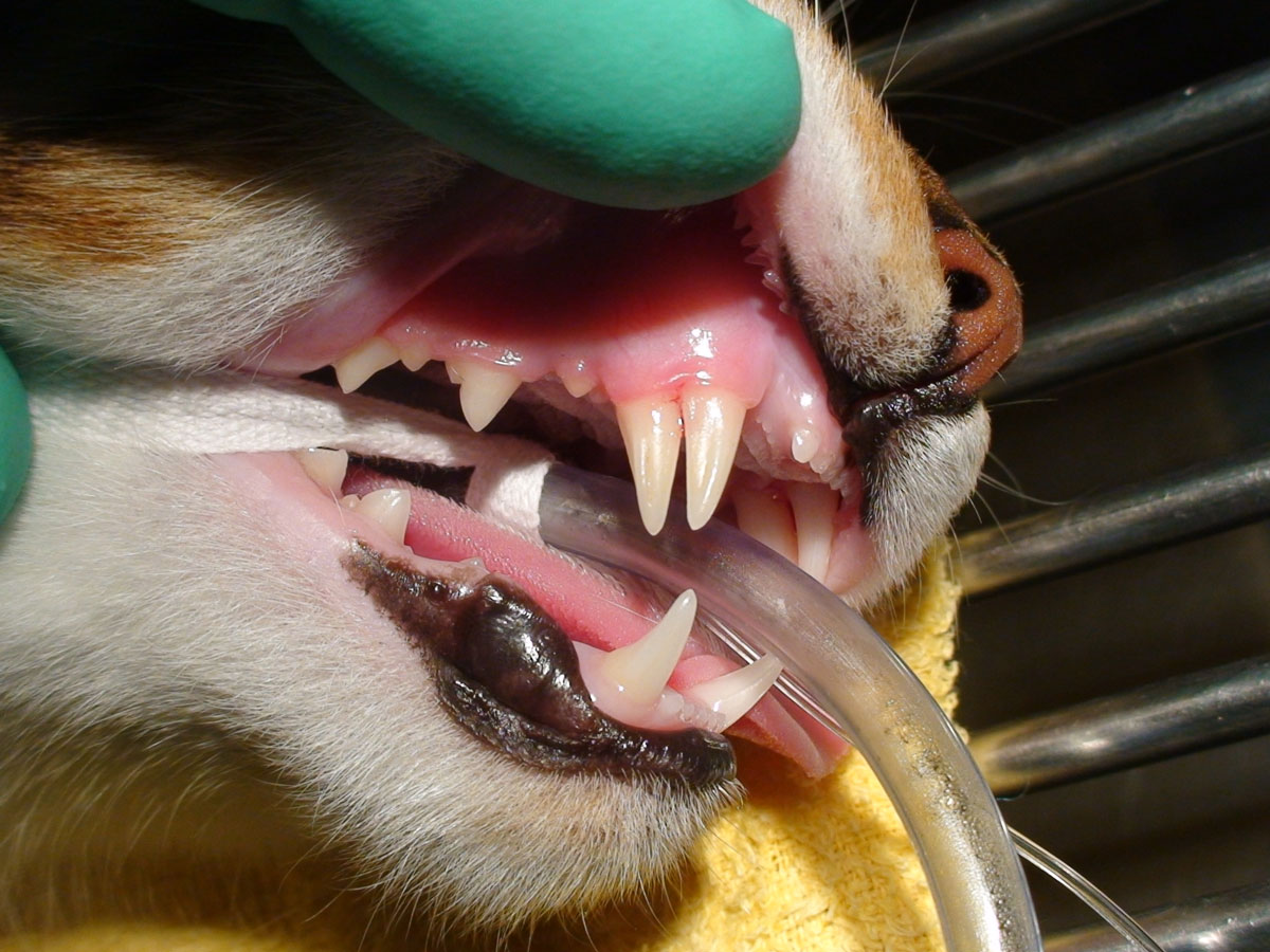
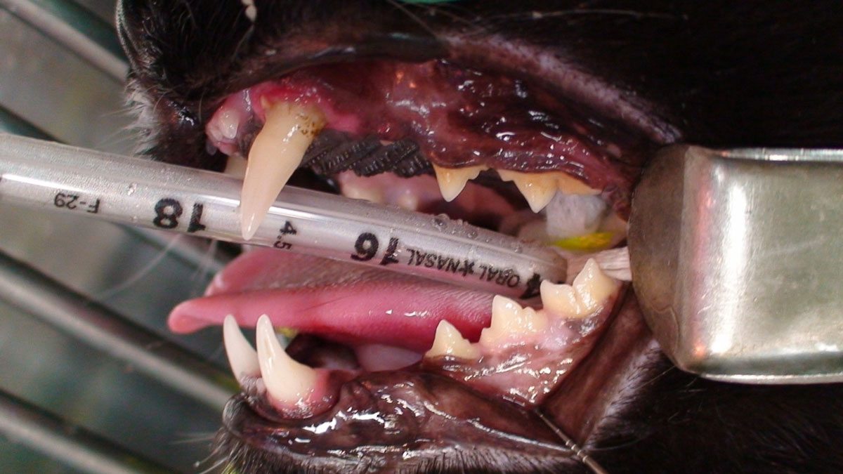
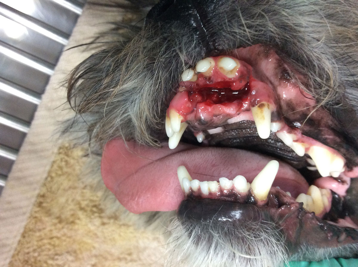
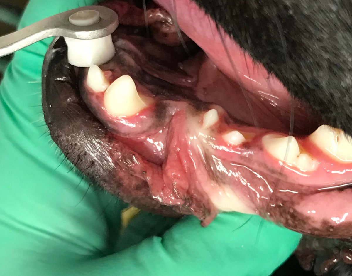
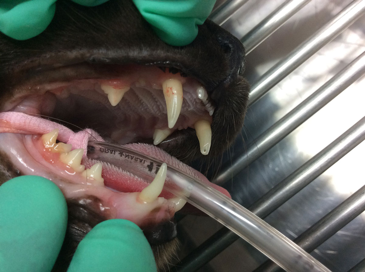
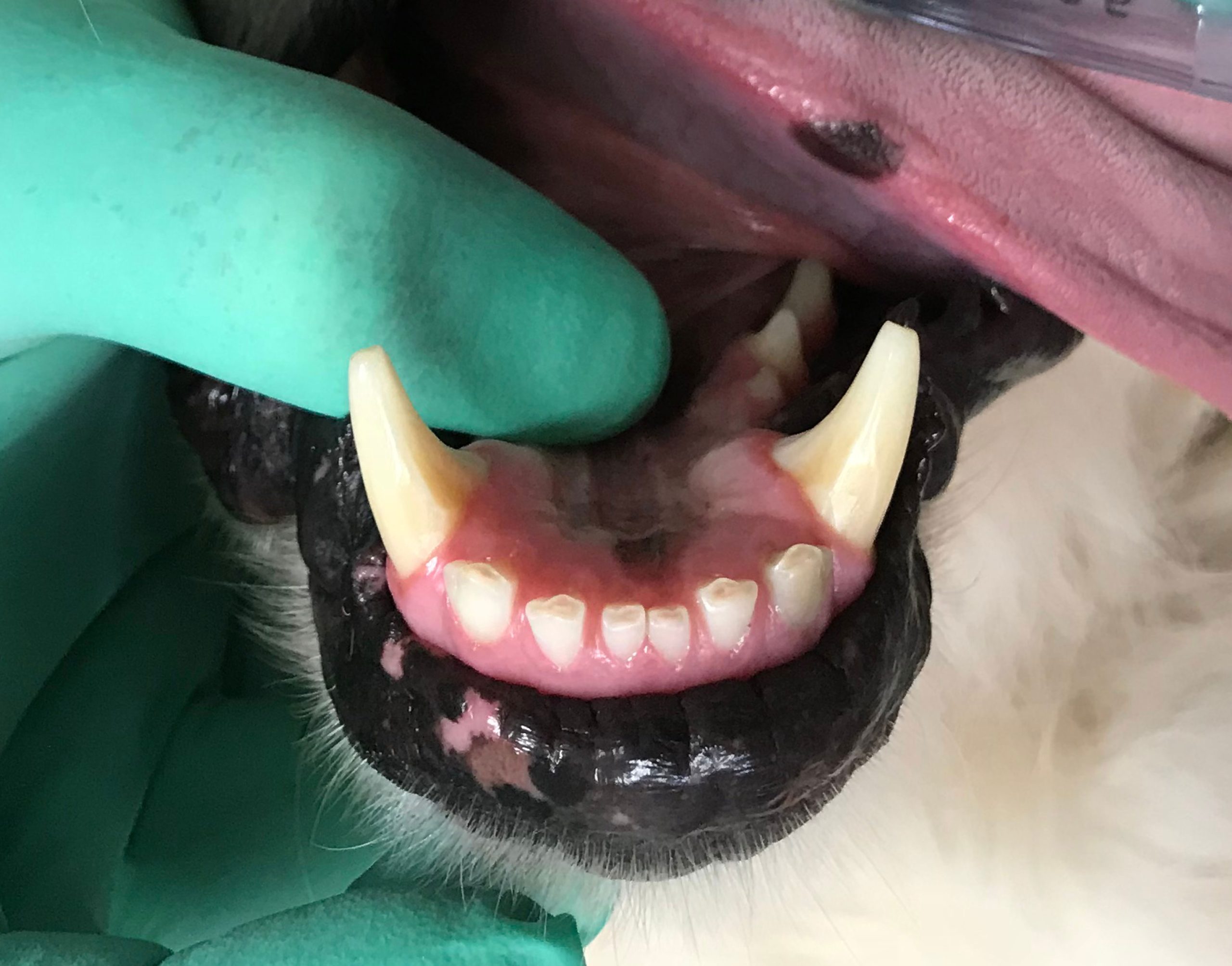
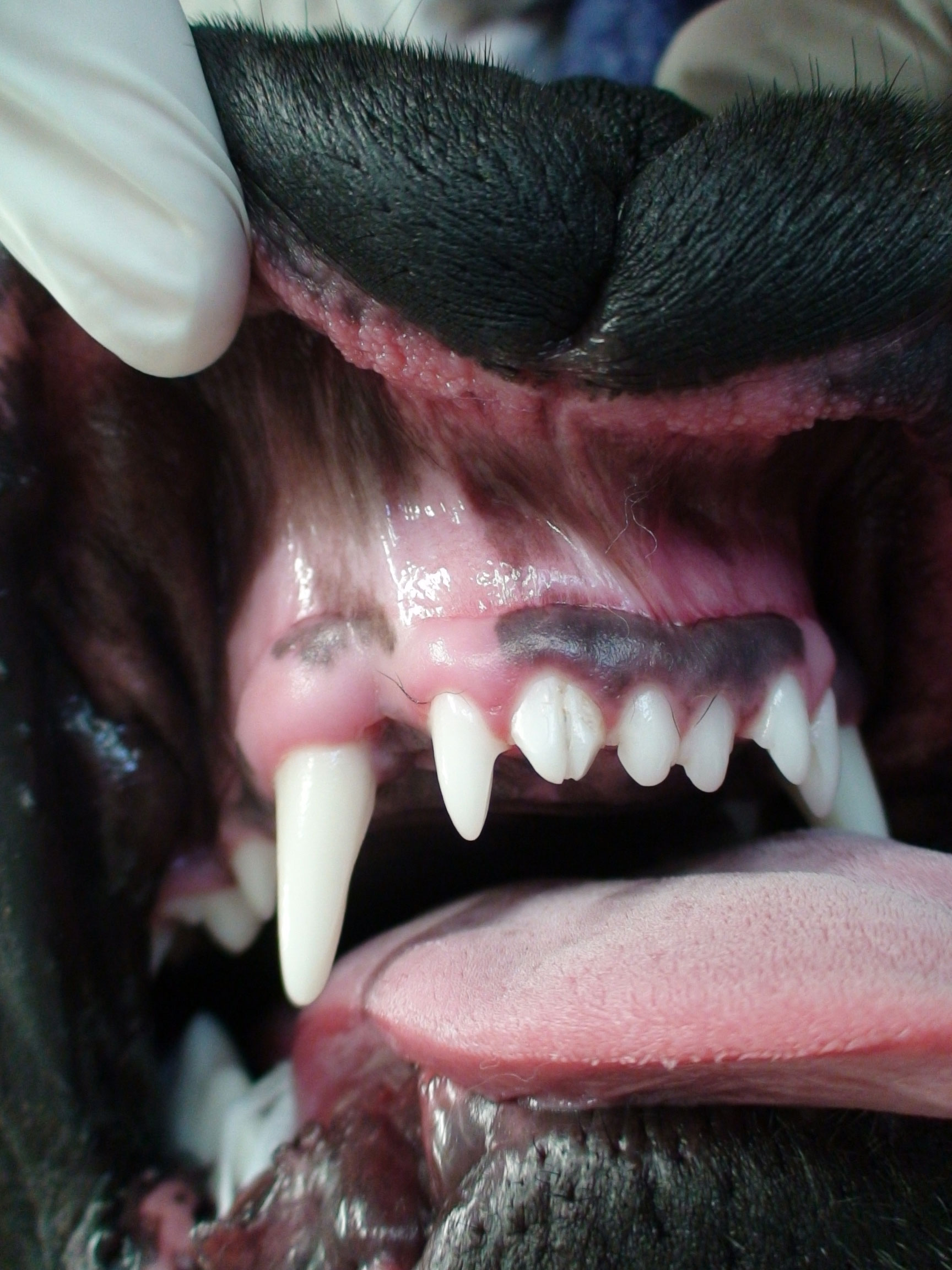
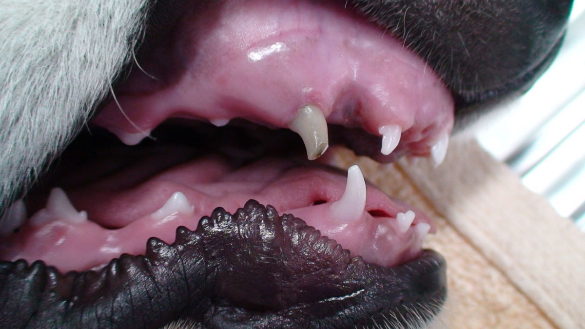
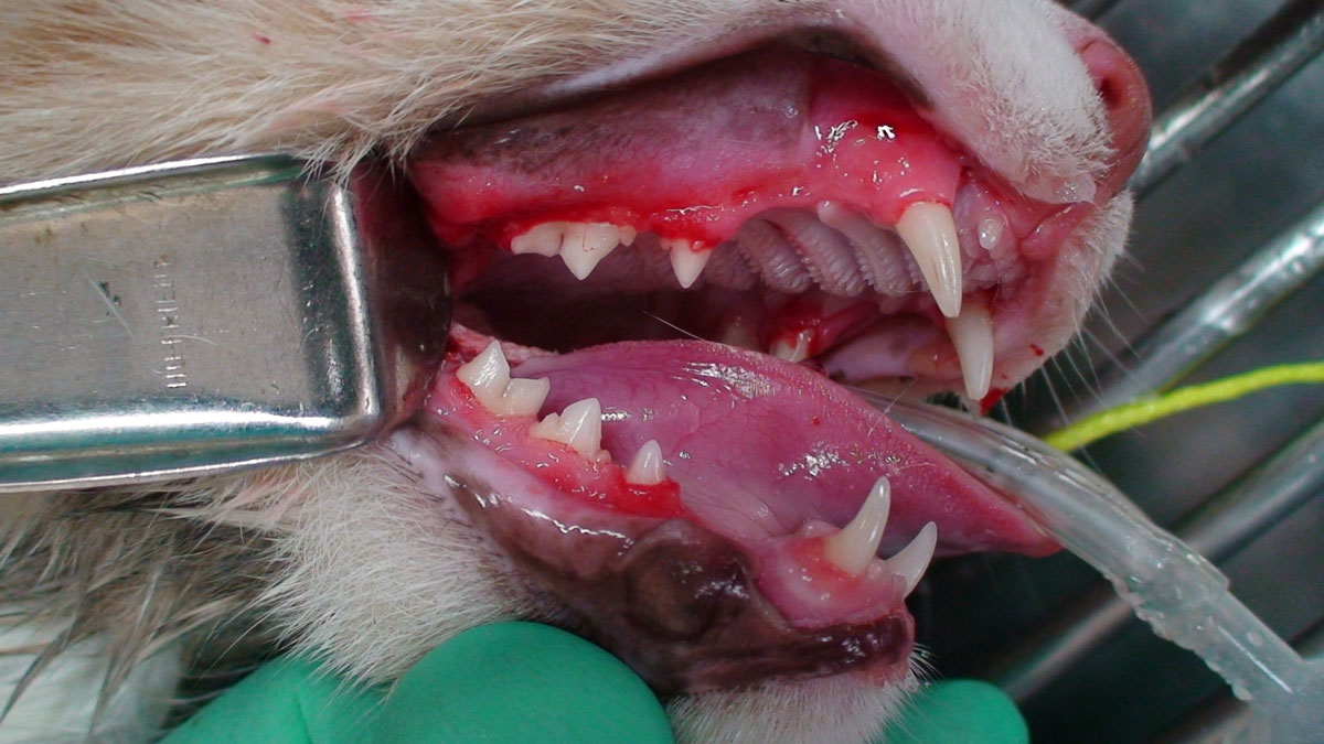
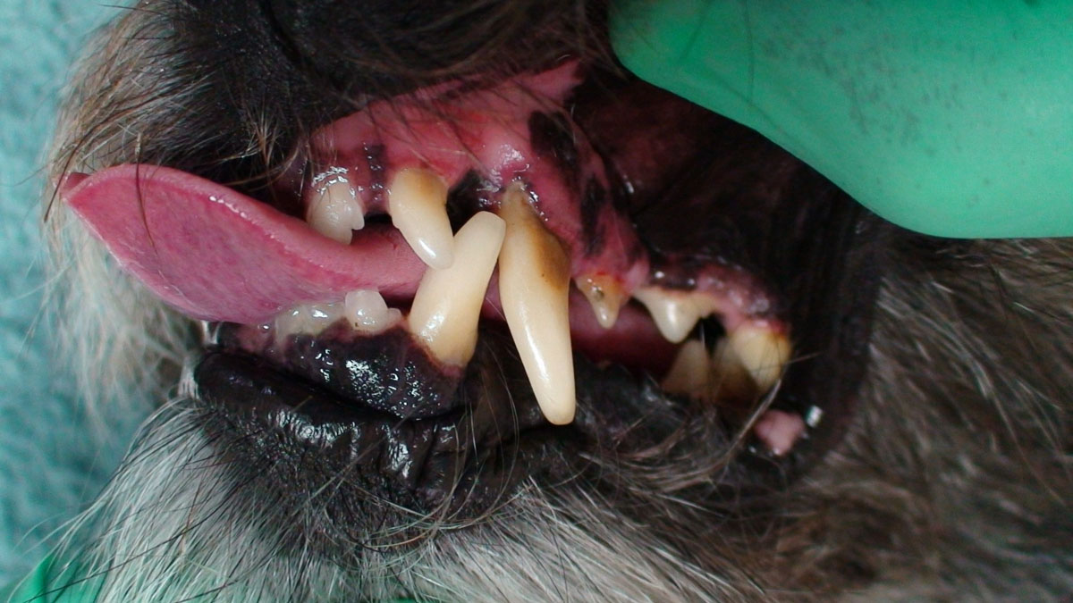
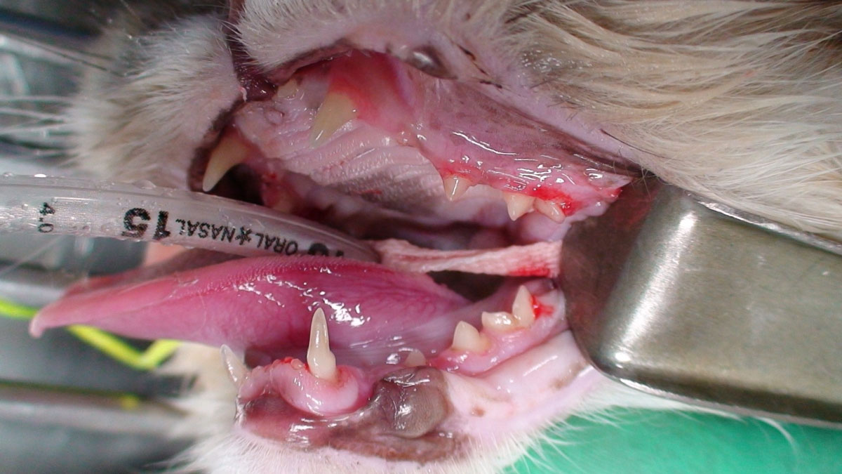
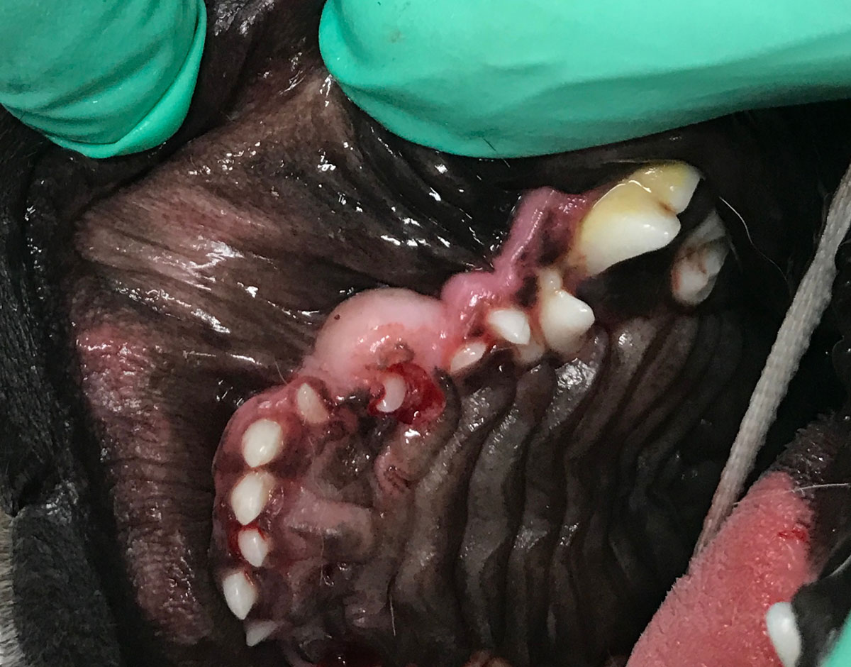
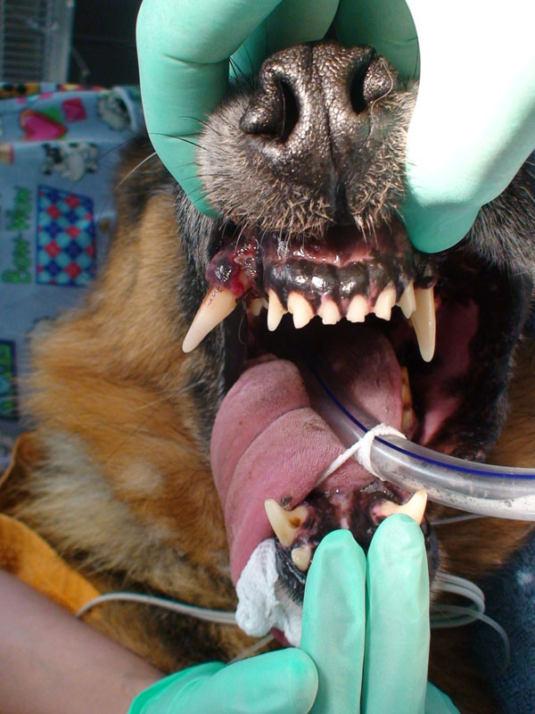
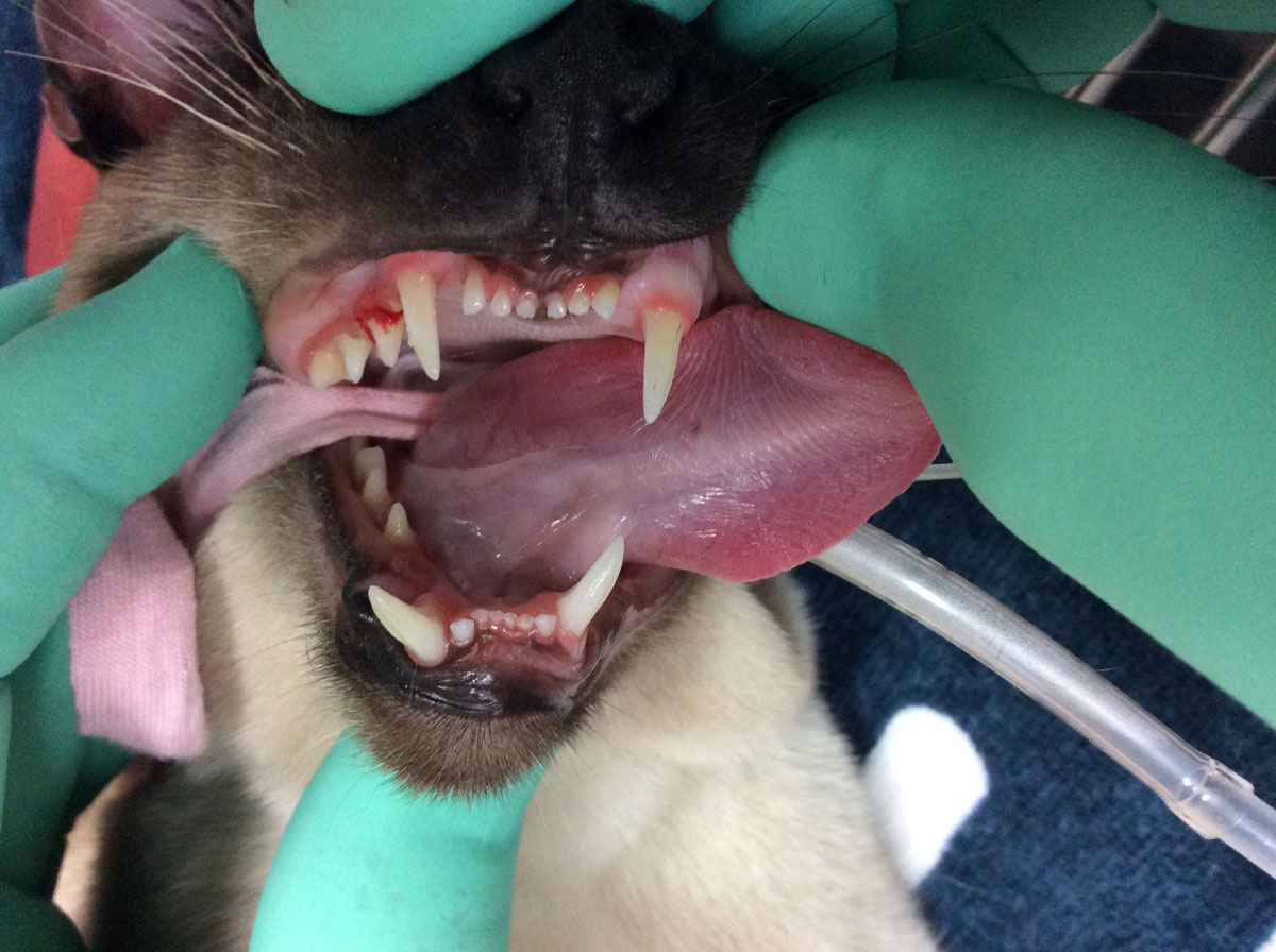
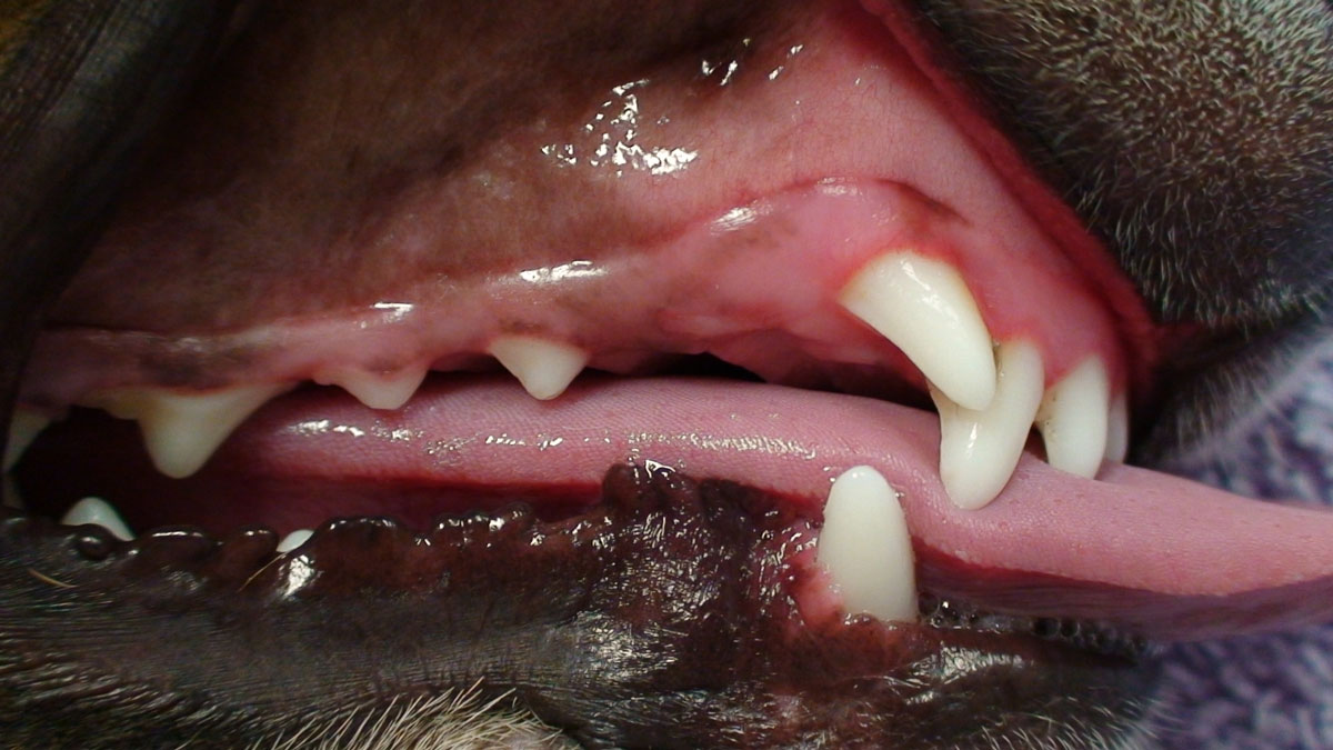
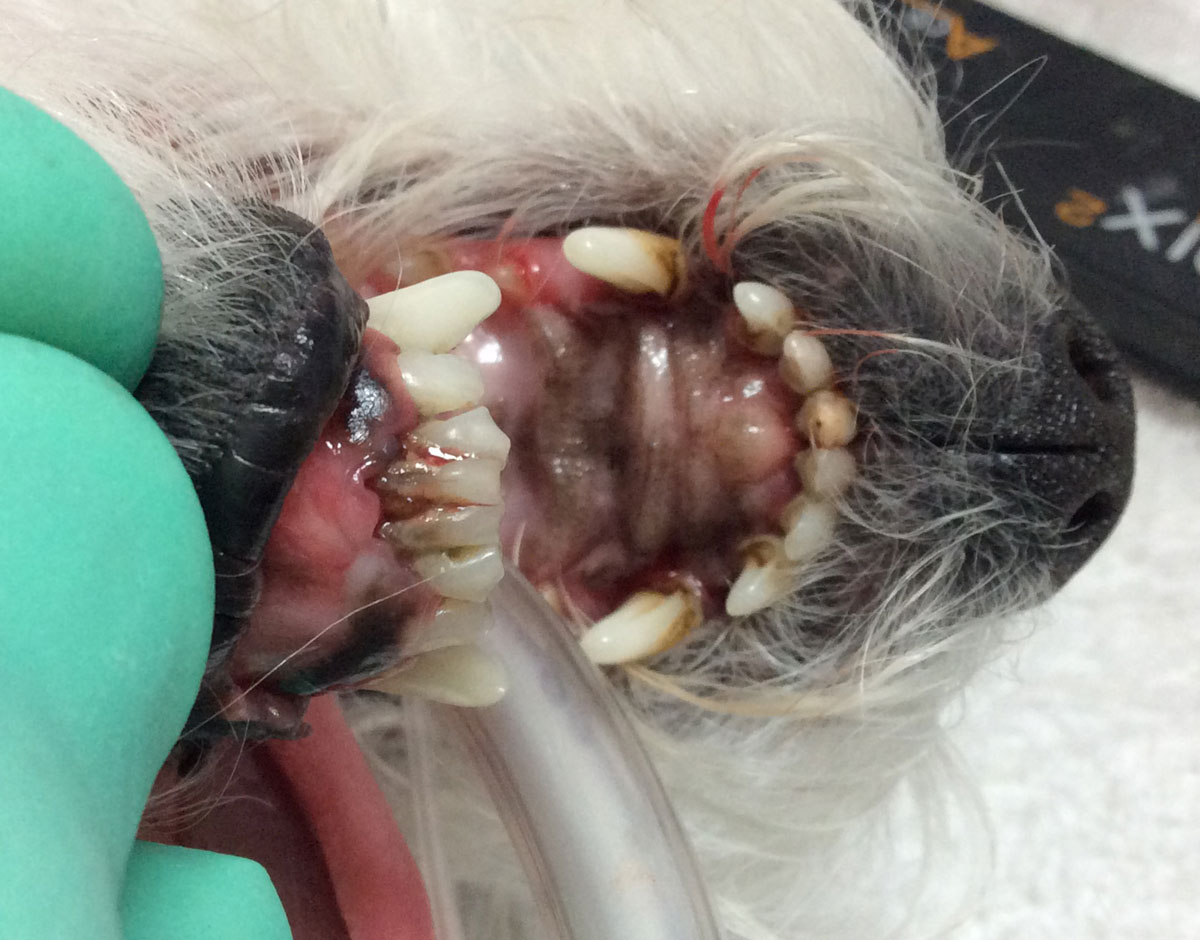
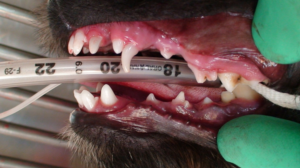
Persistent deciduous tooth
A condition that results in both the deciduous and permanent teeth occupying the same socket due to lack of exfoliation of the deciduous tooth. This often results in the maxillary canine being positioned in a mesial direction, and all other teeth in a palatal or lingual position, resulting in malocclusion.


